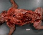
Diaphragmatic Rupture
| This article is still under construction. |
Description
Rupture of the diaphrgam is an acquired condition that often has a traumatic origin in small animals. The rupture is not a true hernia as the parietal peritoneum is disrupted and displaced organs are not contained within a defined hernial sac. Most cases occur in animals which have suffered blunt abdominal trauma with an open glottis, most commonly during a road traffic accident (RTA). If the animal has a closed glottis at the moment of impact, the lung parenchyma is more likely to rupture. Affected animals often have other injuries associated with the traumatic event, including:
- Thoracic injury
- Pulmonary contusion
- Rib fracture or flail chest
- Haemothorax or chylothorax
- Pneumothorax with or without pneumomediastinum
- Tension pneumothorax
- Traumatic myocarditis
- Ruptured trachea
- Abdominal injury
- Ruptured liver or spleen with haemabdomen
- Ruptured kidney, ureter or bladder with uroabdomen
- Traumatic abdominal wall rupture
- Pancreatitis
- Broken claws
- Fractured mandibular symphysis
- Pelvic, spinal or appendicular fractures
- Tail pull injuries
- Wounds
Diagnosis
Clinical Signs
- The animal may have a history consistent with blunt trauma (such as a road traffic accident, kick or blow to the abdomen) and broken claws are a common finding after an RTA.
- Respiratory distress as displaced abdominal organs in the thorax prevent the lungs from expanding fully and because the damaged diaphragm is incapable of contracting normally. Affected animals may also develop pleural effusion if abdominal organs become incarcerated or strangulated in the thorax.
- Heart sounds may be muffled on auscultation and borborygmi may be heard.
- Percussion of the chest wall may reveal hyporeseonance (due to displaced gas-filled stomach) or hyperresonance (due to the presence of pleural fluid or solid organs, such as the liver, in the chest).
- The apex beat of the heart can usually be palpated and this may be displaced from the normal position on the left cranial ventral chest wall.
- In chronically affected animals, gastro-intestinal signs may be observed due to partial intestinal obstruction or pancreatitis.
Radiography
Plain chest radiographs will show that the margin of the diaphragm is no longer evident and abdominal organs, particularly gas-filled loops of small intestine, may be evident within the chest. This appearance should be distinguished from that of peritoneopericardial diaphragmatic hernia in which abdominal organs only overly the cardiac silhouette. If the diagnosis is not certain, a barium swallow series could be performed or contrast medium could be instilled into the peritoneal cavity but these procedures have largely been superseded by the use of ultrasound.
Ultrasound
This technique has been shown to be much more accurate than radiography for the diagnosis of diaphragmatic rupture but care should be taken not to confuse the appearance of displaced abdominal organs with a reverberation artefact generated by the intact diaphragm.
Pathology
Displaced organs begin to form fibrinous adhesions within the chest almost immediately and these organise into fibrous structures over the following seven days. The rent in the diaphragm usually occurs in the muscular portion in small animals, often at different locations depending on the species affected:
- Dog: Either radial or circumferential.
- Cat: Circumferential tears are much more common than radial.
- Horse: Tears usually occur through the tendinous portion.
The edges of the tear are gradually replaced by fibrous tissue and these may need to be resected if a surgical repair is subsequently attempted.
Treatment
Stabilisation
In most acute cases, animals must be stabilised before the tear in the diaphragm can be repaired surgically. This may involve the following steps:
- Provision of oxygen to dyspnoeic animals, using a mask, flow-by or intra-nasal catheter.
- Pleurocentesis if pleural effusion or pneumothorax are suspected.
- Keeping the animal in sternal recumbency to allow more efficient thoracic excursion.
- Gastric decompression by orogastric tube or percutaneously if the stomach is though to be dilated.
- Provision of analgesia.
- Other measures to treat other traumatic injuries.
Surgical Repair
Traditionally, it was recommended that 24 hours elapse from the traumatic event until the rupture was repaired to reduce perioperative mortality but newer evidence suggests that, if animals are adequately stabilised before this, surgical repair may still be successful. If possible, the repair should be conducted in the first week after rupture as fibrous adhesions begin to form after this time. Post-operative mortality is also higher if the rupture is repaired after a very long interval (more than 1 year) due to the formation of extensive fibrous adhesions.
The defect is approached by a ventral midline coeliotomy (which may be extended cranially beside the xiphisternum or into a median sternotomy) and the abdominal organs are retracted. Fibrinous adhesions can be easily separated but strangulated organs (such as torsed liver lobes or loops of small intestine) should be resected. If the rupture has been present for a long period, its fibrous edges may be debrided before suturing using polydioxanone. If there is a large defect that cannot be closed without tension, the following approaches may be used:
- Use of the muscle transversus abdominis as a flap to fill the defect
- Use of porcine intestinal submucosa or synthetic mesh to fill the defect
- Graft of omentum over sutures placed in the diaphragm
Post-operative Care
Animals that have been treated for diaphragmatic rupture often require intensive care in a dedicated unit. The following aspects of care should be considered:
- Thoracostomy tube: This can be placed through the diaphragm during surgery or a conventional tube can be passed through the chest wall. Negative pressure should restored to the pleural cavity after the rupture is repaired and the diaphragm should be seen to return to its concave shape when viewed from the abdomen. The chest should be drained (of air or fluid) regularly until no more can be aspirated than is expected due to the presence of the tube (~2 ml/kg/hour). Bupivacaine or another local anaesthetic agent can be instilled through the tube to provide topical analgesia.
- Pulmonary oedema may develop as the lungs re-expand due to physical forces acting on the alveoli and due to reperfusion injury to the alveolar capillaries. This condition should be suspected if the patient remains hypoxaemic even with oxygen therapy and it can be avoided by removing air from the pleural space slowly so that the lungs reinflate in a controlled manner.
- Incomplete repair or presence of a second rupture: The whole diaphragm should be assessed before closure to ensure that a second tear is not present.
- Provision of analgesia.
Prognosis
Patients that undergo surgical repair of a rupture have a favourable prognosis, with around 90% being discharged after treatment.
References
| Also known as: | Acquired Diaphragmatic Hernia Displacement of Stomach into Thorax |
Do not confuse with: | Hiatal Hernia Peritoneopericardial Hernia |
