Pregnancy - Donkey
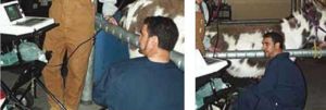
Ultrasonographic characteristics of the asinine early pregnancy have been described, and present several similarities with the horse (Meira et al, 1998; Carluccio et al, 2004).
Early pregnancy diagnosis

The embryonic vesicle can be detected by transrectal ultrasonography from days 10 to 12 post-ovulation (11.5 ± 0.9 days). Fixation of the embryonic vesicle occurs 15.5 ± 1.4 days after ovulation. The mean growth rate of the embryo is 3 to 3.5 mm per day until 18 days. The vesicle becomes irregular and grows more slowly (0.4 to 0.5 mm per day) from day 19 until day 29. The heartbeat is detected at 23.5 ±1.3 days.
Secondary corpora lutea are formed between days 38 and 46 (41.8 ±1).Plasma progesterone increases in the first 19 days to about 19.9 ng/ml then decreases to 12 ng/ml at day 30. Another increase is seen between day 30 to 40 (17 ng/ml). Progesterone gradually declines between 110 and 160 days and remains at about 6 ng/ml. Chorionic gonadotropin is secreted after day 36 of pregnancy (Meira et al, 1998a; Meira et al, 1998b).
Foetal well-being and the foeto-placental unit can be evaluated in the same manner as for the horse. In late pregnancy, the foetus is generally very deep and cranially situated in the abdomen.
Literature Search
Use these links to find recent scientific publications via CAB Abstracts (log in required unless accessing from a subscribing organisation).
Donkey Pregnancy publications
References
- Tibary, A., Sghiri, A. & Bakkoury, M. (2008) Reproduction In Svendsen, E.D., Duncan, J. and Hadrill, D. (2008) The Professional Handbook of the Donkey, 4th edition, Whittet Books, Chapter 17
- Carluccio, A., Villani, M., Contri, A., Tosi, U., and Veronesi, M.C. (2004).‘Ultrasonographic evaluation of the early pregnancy in Martina Franca jennies’. Ippologia 16. pp 31-35.
- Meira, C., Ferreira, J.C.P. , Papa, F.O., and Henry, M. (1998). ‘Ovarian activity and plasma concentrations of progesterone and estradiol during pregnancy in jennies’. Theriogenology 49. pp 1465-1473.
- Meira, C., Ferreira, J.C.P., Papa, F.O., and Henry, M. (1998). ‘Ultrasonographic evaluation of the conceptus from 10 days to 60 of pregnancy in jennies’. Theriogenology 49. pp 1475-1482.
|
|
This page was sponsored and content provided by THE DONKEY SANCTUARY |
|---|
