Introduction
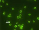
Immunofluorescence is a technique used to detect cell or tissue-associated antigens using antibodies labelled with fluorescent tags. The stained tissues are then detected by immunofluorescence microscopy (qualitative) or flow cytometry (quantitative). Antibodies bind stably and specifically to their corresponding antigen and the technique makes use of the fact that they can be coupled to fluorescent dyes, such as fluorescein and rhodamine, with no effect on specificity. These conjugates bind to antigens present in a sample and can then be visualised under a microscope with a suitable light source, such as UV light. Conversely, the technique can also be used to detect antibodies directed against antigens known to exist in a sample.
Fluorescent dyes
If a molecule has the property of fluorescence, it can absorb light of a one wavelength (excititation) and emit light of another (emission). Antibodies tagged with these dyes (known as fluorochromes) form immune complexes with specific antigens that can be detected when excited by light of a certain wavelength.
Commonly used fluorochromes
- Fluorescein: organic (carbon-based) dye, most widely used. Absorbs blue (490nm) and emits yellow green fluorescence(517nm)
- Rhodamine: organic dye, absorbs yellow-green light and emits deep red fluorescence (546nm).
- As fluorescein and rhodamine fluorescences are easy to distinguish from one another, it is possible to conjugate them to different antibodies and simultaneously visualise two different antigens on the same cell or tissue.
- Phycoerythrin: can absorb light from the blue-green (495nm) and the yellow wavelengths, emits bright red fluorescence
Techniques
Direct staining
- An antibody directed against a specific antigen is directly conjugated with the fluorescent dye and applied to the sample.
Indirect staining
- Utilizes a double layer technique- a primary, unlabelled antibody is applied to the sample, followed by a secondary antibody, an anti-immunoglobulin that has been conjugated to a fluorochrome.
- Indirect staining has several advantages:
- Several secondary antibodies bind to each primary antibody, so the resulting fluorescence is brighter than that of the direct staining.
- One preparation of secondary antibody can be used to test many sera
- By using a mixture of primary antibodies, it is possible to detect the relative expressions of different antigens in the same cell
- Indirect staining has several advantages:
