| This article has been peer reviewed but is awaiting expert review. If you would like to help with this, please see more information about expert reviewing. |
|
|
Introduction
Poultry coccidiosis is caused by Eimeria species. The poultry industry has grown to its massive size through the development and administration of anticoccidial drugs. Most growing birds are fed compounded rations containing anticoccidial drugs which has radically reduced the deaths from coccidiosis bringing the mortality rate down to negligable levels. Careful management is needed to prevent decreased productivity from infection through decreased egg production, weight gain and feed conversion ratios.
Mammalian coccidiosis is usually associated with watery diarrhoea, dysentry and weight loss. It usually presents in young animals which are living in overcrowded and unhygienic conditions.
Pathogenicity of coccidiosis is related to the size of the endogenous stages, the location of the infection in the mucosa and the site of infection. For example, infection in the small intestine can lead to compensation whereas infection in the large intestine will affect water absorption. If mucosal stem cells are affected, it will cause villous atrophy and a slow recovery.
Eimeria spp.
- Many species occur
- All species are host specific
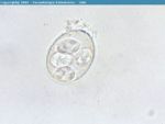
- Sporulated oocyst has 4 sporocysts which each contain 2 sporozoites
- Oocyst measures 20-50μm
- Direct life cycle
- Transmission by the oral-faecal route
Isospora spp.
- Many species occur
- All species are host specific
- Sporulated oocyst conatins 2 sporocysts each with 4 sporozoites
- Oocyst measures 20-50μm
- Usually direct life cycle
- Transmission via the oral-faecal route
- Some species use facultative intermediate hosts and form tissue cysts
- Transmission via faecal-oral route or by ingestion of the intermediate host
Coccidia of Poultry
- Direct life cycle
- 1 week prepatent period
- After oocysts are ingested, sporozoites are released which penetrate the intestinal epithelium
- 2 asexual phases of multiplication called schizongony occur followed by a phases of sexual multiplication called gametogeny
- Zygote develops an oocyst which is shed in the faeces
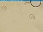
- Oocyst measures 20-30μm
- For each oocyst ingested, thousands are shed
- Life cycle is self-limiting
- Organisms from a single infection go through the sequence of developmental stages synchronously
- Organisms leave the body simultaneously as oocysts
- Oocysts are only infective once they have sporulated
- Sporulation requires warmth, moisture and oxygen
- Takes 2-3 days in broiler houses
- Oocysts contain 4 sporocysts each with 2 sporozoites
Pathogenesis
- 7 important Eimeria species
- 4 malabsorptive species
- Eimeria acervulina which is moderately pathogenic
- Eimeria maxima which is moderately pathogenic
- Eimeria mitis which has low pathogenicity
- Eimeria praecox which has low pathogenicity
- 3 haemorrhagic species
- Eimeria tenella
- Eimeria necatrix
- Eimeria brunetti
- All highly pathogenic
- Form large sub-epithelial second generation schizontsat the base of intestinal crypts
- Deep eruptions form when cells rupture to release merozoites
- Destruction of crypt stem cells and marked haemorrhage
- Blood stained faeces
- High morbidity and high mortality
Diagnosis
- Post-mortem diagnosis of lesion severity
- Region of intestine affected
- Appearance of lesion
- Presence or adsence of haemorrhage
- Size of schizonts and oocysts found in mucosal scrapings
- Eimeria acervulina
- Proximal gut
- Thickening of walls
- 'White ladder lesions' produced by dense foci of gamonts and oocysts
- Watery exudate
- Eimeria maxima
- Mid-gut
- Thickening of walls
- Pink exudate
- Eimeria tenella
- Swollen caeca
- Thickening of wall
- Dark colouring containing a core of necrotic tissue and blood
- Lesion scoring is the best method of diagnosing the severity of the lesions and therefore the causative Eimeria species
- Eimeria necatrix
- Mid-gut
- Ballooning of wall
- White spots and petechiae forming 'salt and pepper' lesions
- Haemorrhage into lumen
Immunity
- Different Eimeria species produce different levels of protective immunity
- E.maxima -> E.brunetti and E.acervulina -> E.tenella and E.necatrix
- There is no cross immunity between species
- There is very little passive immunity
- Evokes a cell-mediated response
- All ages of poultry are susceptible
Epidemiology
- Oocysts are ubiquitous and robust
- Able to survive several months to years
- It is impossible to keep buildings free from infection
- Chicks become infected by pecking the ground shortly after being placed in the poultry house
- Biotic potential is enormous
- Generation time is short
- Massive infections can build up rapidly
- Immunity develops relatively slowly
- With high stocking densities the situation is explosive
Control
- Chemical
- Intensive poultry production is largely dependent on the use of anticoccidial drugs
- For more information see here
- Vaccines
- Paracox
- Multivalent attenuated live vaccine for replcement layers and broilers
- Contains 7 live strains of Eimeria
- Lack the most pathogenic life cycle stage making the prepatent period shorter
- Known as precocious strains
- Chicks vaccinated on a single occasion when 1-9 days old through oocyst suspension in the feed or water
- Vaccinated birds have sub-optimal growth rates so is not used for broilers
- Paracox 5
- Contains 5 strains of the most pathogenic Eimeria
- Used for broilers
- Sprayed onto the first feed offered to new batches of chicks
- Paracox
- Integrated control
- Careful management is needed so in-feed prophylaxis and vaccination do not fail
- Remove litter and thoroughly clean houses in between crops
- Optimum turn-around time is 10 days
- Use the lowest stocking density which is compatible with economic production
- Water bowls, roofs and walls should be well maintained to prevent litter becoming damp
- Stress factors should be avoided and adequate nutrition provided
Other Avian Coccidia
Coccidia of Turkeys
- 5 Eimeria species
- 2 important pathogenically
- Eimeria in the anterior and mid-intestine causes necrotic enteritis and petechial haemorrhages
- Causes watery diarrhoea in young poults and some mortality
Coccidia of Geese
- 3 Eimeria species
- 2 intestinal species causing macroscopic lesions in kidney tubules
- Oocysts carried in urine and pass out with faeces
- Renal species cause severe disease in goslings
- Depression, emaciation, diarrhoea and sometimes death
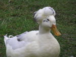
Coccidia of Ducks
- Several Eimeria species
- Another coccidia species which produces 8 sporozoites which are not enclosed in a sporocyst
- Causes severe enteritis and mortality in ducklings
- Haemorrhages and pale focal lesions in small intestine
Coccidia of Game Birds
- 3 main species
Coccidia of Cattle
- Many species affect cattle
- Cattle under a year old are usually infected sporadically
- 2-3 week prepatent period
- Eimeria bovis
- Eimeria zuernii
- Endogenous stages in connective tissue of lamina propria of the lower small intestine and in the epithelial cells of the caecum and colon
- More pathogenic than Eimeria bovis
- Causes blood stained dysentry, tenesmus and sloughed mucosa
- Oocysts are spherical and measure 16μm
- Mainly occurs in calves in poor conditions and brought-in calves
- Also occurs in suckler calves turned out in spring
- Eimeria alabamensis associated with diarrhoea in calves after spring turnout
- Passive immunity is sufficient during the neonatal period
- Can be concurrent with cryptosporidium, viral and bacterial agents
Diagnosis
- History, clinical signs, diarrhoea (often with blood) and a decrease in weight gain
- Post-mortem
- High faecal oocyst count
- However, healthy animals can pass millions of oocysts from mixed species infections which have no pathogenic significance
- Animals may die before oocysts are shed
Control
- Improve husbandry
- Improve sanitation
- Increase bedding
- Raise faecal and water troughs to avoid faecal contamination
- Preventative in-feed medication
- E.g. Decoquinate
- Injectible antiprotozoals may limit oocyst production but animals should still be moved to a clean environment
- E.g. Sulphamethoxypyridazine
Coccidia of Sheep
- 11 different Coccidia species although only two are of clinical significance
- Giant schizonts visible as white spots
- Eimeria crandalis
- Varying pathogenicity
- Scours, grey, foul-smelling faeces
- Parasitises the small intestine, caecum and colon
- 2 week prepatent period
- Disease frequently seen in lambs under 6 months old
- More often in twins and triplets when single lambs
- Oocyts from ewes (immune carriers) accumulate in poorly managed litter or around feed and water troughs
- Lambs born early in the year amplify the parasite problem increasing the parasite risk to lambs born later in the year
- Affected lambs may die before oocysts are found in the faeces
- Post-mortem diagnosis difficult
- Different species of Eimeria occurs in sheep and goats
- Infection may be coincident with Neospora or Cryptosporidium infections
- Mixed infections complicate the diagnosis as oocyst differentiation is difficult
- Other non-pathogenic species can cause papillomatous mucosal growths
Control
- Improve husbandry
- Avoid overcrowding
- Decrease stress
- Improve hygiene by dagging ewes
- Avoid mixing lambs of different ages
- Preventative measures include creep feeding lambs with decoquinate or oral dosing with diclazuril when lambs are 4-6 weeks
- A second dose can be given after 3 weeks
Coccidia of Goats
- Many Eimeria species
- 2 Eimeria are pathogenic
- Cause diarrhoea and a decreased growth rate
- Different species of Eimeria occurs in sheep and goats
Coccidia of Horses
- Only one atypical Eiemria
- Forms large subepithelial gametocytes in villi
- Large, dark coloured oocysts
- Approximately 12μm
- Occasionally causes diarrhoea
- Besnoitia bennetti in larynx of horses
Coccidia of Pigs
- Many species of Eimeria and Isospora
- Only Isospora suis is of clinical pathogenic importance
- Causes sporadic, serious and sometimes fatal disease in unweaned piglets
- Causes profuse diarrhoea
- Very short 1 week prepatent period
- Diarrhoea starts before oocysts are shed in faeces
- Ante-mortem diagnosis is difficult
- Death usually occurs after parasites have left the host
- Post-mortem diagnosis difficult
- Isospora infections are self-limiting
Coccidia of Dogs
- 2 common and 2 less common Isospora species
- Occasionally can cause disease
- Little pathogenicity
- Even if faecal oocyst count is high, other causes of diarrhoea should be looked for
- Hepatozoon americanum and subclinical H. canis in periosteal bone formation
- Both are Tick borne diseases
- H. canis – Rhipicephalus sanguineus
- Ticks become infected by ingesting a blood meal containing macrophages and neutrophils infected with the parasite gamonts -> sexual replication in the gut of the tick -> oocysts containing infective sporozoites -> dogs ingest the tick schizogony occurs in numerous tissues
- Both are Tick borne diseases
Coccidia of Cats
- 2 common Isospora species with little clinical significance
- Oocysts in faeces have to be distinguised from those of Toxoplasma (smaller) and Sarcocytis (sporulated or naked sporocyts in faeces)
Coccidia of Rabbits
- 3 pathogenic Eimeria species
- 2 in the caecum
- 1 in the bile duct
- Eimeria steidae
- Serious disease of both pet and farmed rabbits
- Treatment is by administration of drugs in drinking water
- E.g. Toltrazuril
- Hygiene is the best method of prevention to prevent sporocysts from sporulating
- Medicated feed can be used in commercial units
- E.g. Rabenidine
