| This article is still under construction. |
Overview
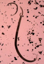
The structure of a nematode is intimately related to its function and its life cycle. Although there are common traits throughout the phylum there is also great diversity allowing each species to occupy a niche in which it may thrive.
Body Structure
The nematode body is cylindrical, elongated and smooth with no limbs protruding, such as is seen in the common garden worm though generally on a smaller scale. The body is contained within a tough elastic cuticle which in many species forms elaborate structures useful for identification. The presence of a cuticle is similar to the structure of arthropods, however unlike them the nematode cuticle is not chitinous but is comprised mainly of collagens. The cuticle is non-living, produced by cells of the epidermis in most of the worm, allowing it to grown between moults of the worm without the need for shedding, although this does occur a number of times during the development of most worms. It is permeable to allow ions and water to pass through and therefore plays a key role in maintaining the hydrostatic pressure, which in most nematodes is relatively high inside the worm. The cuticle also acts as an anchoring point during locomotion as a skeleton does in mammalian species.
Morphological differences in the cuticle are regularly used to identify different species of nematodes, though the functions of these are not all completely understood.
- Annulations - Transverse lines in cuticle, possibly used as anchoring points in locomotion
- Longitudinal Ridges - also known as 'synlophe', seen in some Trichostrongylidae species such as Nematodirus.
- Alae or wings - Projections of the outer cuticle layer. Can appear either just at the anterior or posterior or along the entire length of the worm. In bursate males posterior alae for part of the copulatory bursa.
- Spines - Protrusions of the cuticle on the surface of the nematode. Function unknown, could be in self defence or attachment to host.
- Inflations - Vesicle like swellings of the cuticle function unknown. Found in Oesophagostomum species.
Locomotion
Locomotion in nematodes involves somatic muscles that are present below the cuticle and hypodermis. They are attached to the hypodermis and separated into four sections by hypodermal cords. They are obliquely striated unlike mammalian muscles and have dense bodies as opposed to Z disks. Two types of muscle arrangement occur in nematodes, platymyarian in small worms and coelomyarian in larger worms. During locomotion the muscles are used to apply pressure laterally to the cuticle, this pressure is opposed by the high hydrostatic pressure of the coelom and causes dorso-ventral bending. These muscular contractions cause the nematode moves in a 'sinusoidal' manner.
Nervous System
Knowledge of the nervous system employed by nematodes has enabled the development of many anti-parasitic drugs as they work to disrupt this system. There is a neural ring around the pharynx of the nematode containing 4 ganglia which communicate distally to the body of the nematode to co-ordinate movement.
Feeding and Digestion
The pharynx is situated at the anterior end of the nematode and is used in feeding, often being embedded into the epidermis or blood vessels of the worms predilection site. The pharynx may be specialized depending on the predeliction site and food type that the nematode requires, many blood feeders have teeth or plates used for attachment. The pharynx has a radial muscle that is used in pumping food into the intestines. The food enters the buccal capsule the size and shape of which is characteristic in some species of nematode. Due to the high pressure levels in the nematode body cavity there is a one way valve between the oesophagus and intestines and food is pushed through this by peristaltic action of radial oesophageal muscles.
Digestion occurs rapidly and faeces is expelled under pressure from the posterior of the nematode.
Reproduction
Recognition Features
- The shape of the pharynx is characteristic in some groups
- There is a nerve ring around the pharynx and four longitudinal nerves with ganglia that co-ordinate movement (many anthelmintics act by disrupting neuromuscular co-ordination)
- The sexes are separate:
- the female tail generally ends in a blunt point
- males usually have two chitinous rods that can be protruded through the cloaca to hold the female - these are called spicules and, being chitinous, are easily seen under the microscope. As these differ in shape and size between species, they are very useful in identification
- The bursate nematodes are characterised by a large expansion of the cuticle of the male tail to form a clasping organ (the bursa)
- The heads of some nematodes have structures such as:
- leaf-like lips around the mouth (the leaf-crown)
- a buccal cavity
- teeth or cutting plates
Feeding Habits
- Many intestinal nematodes are closely applied to the mucosal surface
- Some swallow ingesta and/or host secretions.
- Others suck a plug of mucosa into the buccal cavity (plug feeders), leaving a circular ulcer
- Yet others bury their heads deep into the mucosa and suck blood
