Colic Diagnosis - Clinicopathologic Evaluation
| This article has been peer reviewed but is awaiting expert review. If you would like to help with this, please see more information about expert reviewing. |
Laboratory tests can be performed to assess the cardiovascular and metabolic status of the patient.
- Packed Cell Volume (PCV)
- Total Plasma Protein (TPP)
- Complete Blood Count (CBC)
- Blood Gases
- Electrolyte levels
Repeat PCV, TPP and CBC should be performed in less critical patients as a guide to response to therapy. In more severe or recurrent cases of colic, theses tests should be performed alongside blood gas analysis and electrolyte levels. For normal horse hematology and biochemistry values click here.
Packed Cell Volume and Total Plasma Protein
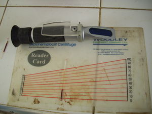
Packed Cell Volume (PCV) and Total Plasma Protein (TPP) are a measure of hydration status in the horse with abdominal pain. Intestinal disease and dysfunction causes hypovolaemia which result in dehydration. Increasing values over repeated examination and values over 45% are considered significant. The total protein (TP) of blood may also be measured, as an aid in estimating the amount of protein loss into the intestine. Its value must be interpreted along with the PCV, to take into account the hydration status. The PCV and TPP rise together in dehydration.
| PCV (%) | TPP (g/dl) | |
|---|---|---|
| Mild dehydration | 45 - 50 | 7.5 - 8.0 |
| Moderate dehydration | 50 - 60 | 8.0 - 9.0 |
| Severe dehydration | 60 | 9.0 |
An increasing PCV without a corresponding rise in TPP may indicate that protein is being lost from the blood into the intestinal lumen or peritoneal region. It may also be due to the spleen contracting and releasing more red blood cells into the circulation in response to endotoxin release or sympathetic nervous system innervation.
Complete Blood Count
A Complete Blood Count can be useful in the colic patient. In cases of acute inflammatory disease such as colitis and enteritis, a leucopaenia (<4000 cells/ul)with a left shift and toxic neutrophils can be seen. Septic peritonitis due to a ruptured intestine will also show a severe leucopaenia (<1000 cells/ul. In chronic peritonitis due to intraabdominal abscesses, a high TPP and high fibrinogen levels alongside a mature neutrophilia will be seen. There are no major changes in the CBC White blood cell count in the early stages of simple and strangulating obstructions. Changes are apparent in the terminal stages.
Blood Gases
A metabolic acidosis with respiratory compensation is a common abnormality seen in colic. Typical values include pH 7.3 units, PCO2 35 mmHg and HCO3- 15 mmol/L. Horses with simple obstructions may show an insignificant base excess compared to those with a strangulating obstruction showing a significant base deficit. It is important to monitor and correct the acid-base imbalances especially for horses that will undergo anethesia and surgery. Rapid deterioration in acid-base status is a poor prognostic indicator.
Serum/Plasma Electrolyte levels
This is the least useful of the hematological tests for establishing a diagnosis in the colic patient, however its importance lies in the management of patients before, during and after surgery.
Hypocalcaemia can cause ileus and abdominal pain. Patients with colitis may have a hyponatremia and hypochloremia. Gastric dilatation results in the sequestration of fluid and hydrochloric acid in the stomach, leading to dehydration, hypochloremia and alkalosis. The same principle applies to large colon obstructions where fluid is trapped in the intestinal lumen causing dehydration and hypochloemia.
Blood and peritoneal lactate levels are useful in determining severity of disease and as a prognostic indicator. Blood levels between 1-2mmol/L are considered normal, while levels above 5.7mmol/L suggest hypoperfusion secondary to dehydration and/or a local ischaemia or strangulating obstruction. Elevated lactate concentrations in the peritoneal fluid are more suggestive of a strangulation.
Horses with dehydration and endotoxaemia may develop a pre-renal azotaemia (increased urea and creatinine).
Increased serum GGT concentrations indicate liver disease. Increased serum bile acids indicate cholestasis. Increased bilirubin levels can be due to anorexia, hemolysis or a hepatopathy.
Muscle damage in horses with severe pain and self-inflicted damage will produce elevations in AST, LDH and creatine phosphokinase.
References
- Edwards B. (2009), Diagnosis and Pathophysiology of Intestinal Obstruction, in Equine Gastroenterology courtesy of the University of Liverpool, pp 9
- Meuller E, Moore J. N, (2008) Classification and Pathophysiology of Colic, Gastrointestinal Emergencies and Other Causes of Colic, in Equine Emergencies- Treatments and Procedures, 3rd Edition, Eds Orsini J. A, Divers T.J, Saunders Elsevier, pp 110 -111
- Rose R.J, Hodgson D.R (2000) Examination of the Alimentary Tract, Alimentary Tract, Manual of Equine Practice, 2nd Edition, Saunders Elsevier, pp 288 -289
