Mandibular Gland - Anatomy & Physiology
Overview
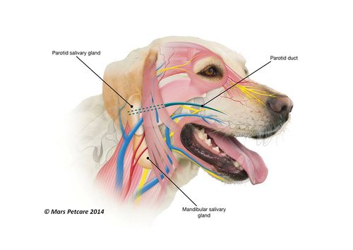
The mandibular gland produces a merocrine secretion, which means it is a mixed gland - serous and mucous secretions. It is generally smaller than the parotid gland and is a moderately large gland in carnivores and herbivores. It is compact and located around the angle of the jaw.
The mandibular duct runs ventral to the mucous membrane of the floor of the oral cavity, close to the frenulum of the tongue. The duct opens at the sublingual caruncle.
Histology
The mandibular gland is known as a tubulo-acinar gland. It consists of mucous cells that stain lighter and serous demilunes that stain darker. Demilunes secrete into lumen by canaliculi between mucous cells.
Innervation
The mandibular gland is innervated by the facial nerve (CN VII) via the chordi tympanii and by the trigeminal nerve (CN V).
Species Differences
Carnivores
The mandibular gland produces mainly mucous secretions in the dog and cat. It is an oval shape in the canid.
Herbivores
The mandibular gland in herbivores is larger and deeper.
| Mandibular Gland - Anatomy & Physiology Learning Resources | |
|---|---|
To reach the Vetstream content, please select |
Canis, Felis, Lapis or Equis |
 Test your knowledge using flashcard type questions |
Salivary Gland Anatomy & Physiology Flashcards |
 Selection of relevant PowerPoint tutorials |
Oral cavity histology tutorial, which looks at salivary glands |
Webinars
Failed to load RSS feed from https://www.thewebinarvet.com/gastroenterology-and-nutrition/webinars/feed: Error parsing XML for RSS
