Ribs and Sternum - Anatomy & Physiology
Costae
Costae are arranged in pairs and articulate with two successive vertebrae. Inidividual ribs have a bony dorsal part, a body of rib, and ventral costal cartilage. They increase in length, curvature and amount of cartilage craniocaudally. The cartilage of last rib may fail to join that of its neighbor and is said to be a floating rib. Costae join ventrally on the midline at the Sternum, which is comprised of three parts, the manubrium, sternebrae and xiphoid cartilage. The manubrium is the most cranial and projects beyond the first set of ribs and can be palpated in most species. The sternebrae is joined by cartilage in young animals that is later replaced by bone and is the main body joining the ribs on the midline. The xiphoid Cartilage is caudal and projects between lower ends of costal arches, providing attachment for the linea alba.
There are a number of costal joints and costosternal joints. The costal joints include the costovertebral joint, where the head of rib articulates with vertebral column via a ball and socket with very restricted mobility and the costotransverse joint where a tubercle articulates with the vertebra via a sliding joint. The costosternal joints include the interchondral joint that is within the rib itself and is an elastic syndesmoses and the intersternal joint which is an impermanent synchondroses between the rib and the sternum.
Thoracic Musculature
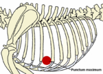
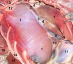
The thoracic muscles are primarily concerned with respiration. Inspiratory muscles enlarge the thoracic cavity whilst expiratory muscles diminish the cavity and force air out. The most important thoracic muscle is the diaphragm, which separates the thoracic and abdominal cavities. It is dome-shaped and convex on its cranial surface. Central tendons form the vertex of the diaphragm. In a neutral position (between full inspiration and full expiration) the diaphragm is located at the 6th rib behind the olecranon. The diaphragm attaches via a muscular periphery to the costal arch.
The diaphragm has three openings; the aortic hilus which conveys the aorta, azygous vein, and thoracic duct, the oesophageal hiatus which conveys the oesophagus, vagal trunks and supplying vessels and the caval foramen within central tendon conveying the caudal vena cava. The diaphragm is innervated by the phrenic nerve, which arises from the caudal cervical nerves (C5-C7).
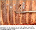
The intercostal muscles are external muscle fibers that run caudoventrally and internal muscle fibers run cranioventrally, although each is confined to a single intercostal space. The transversus thoracis arises from and covers the dorsal sternum and inserts on sternal ribs close to the costochondral junctions. The rectus thoracis covers the ends of the first four ribs in continuation of the rectus abdominus. The serratus dorsalis overlies the dorsal aspect of the ribs. Innervation of the intercostal muscles is supplied by the intercostal nerves, which are ventral branches of the thoracic spinal nerves.
Abdominal Musculature
- Ventrolateral Muscles: flanks and abdominal floor
- All muscles join via aponeuroses in the linea alba at midline, which runs from the xiphoid process to the pelvic symphysis via the prepubic tendon, ensheathing the rectus abdominus
- The External abdominal oblique runs caudoventrally from the lateral surface of the ribs and the lumbar fascia to the linea alba. Its caudal border is thickened to form the inguinal ligament and a slit in its aponeurosis forms the superficial inguinal ring.
- The Internal abdominal oblique runs cranioventrally from the tuber coxae and the thoracolumbar fascia to the linea alba. It forms the cranial border of the inguinal canal.
- The Transversus abdominus is the deepest muscle of the flank, running dorsoventrally from the inner surface of the last ribs and the transverse processes of the lumbar vertebrae.
- The Rectus abdominus forms a broad band parallel to the linea alba, arising from the ventral costal cartilages and inserting on the prepubic tendon. It also forms the medial border of the inguinal canal.
- Sublumbar Muscles:
- Psoas minor: stabilizer of the vertebral column, may also rotate the pelvis at the sacroiliac joint
- Psoas major and Iliacus:


