Introduction
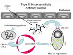
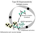
In the normal animal, immune complexes (lattice of soluble antigen and antibodies) are formed and removed all the time. They are broken up by complement, transported to the spleen by RBC's and phagocytosed. When the amount of immune complexes formed (due to rapid influx of antigen) does not equal the amount that are being cleared it causes type III hypersensitivity (an excess of immune complexes).
Location of the immune complexes:
Locally:
1. Inhaled antigen leads to hypersensitivity pneumonitis
- Farmers lung (humans) - inhalation of fungal spores
- Pigeon fancier's disease - repeated inhalation of dried pigeon faeces
- Mouldy hay containing Micropolyspora felis
2. Intradermal and subcutaneous injection of antigen (with high levels of circulating antibody) leads to localised immune complexes which cause acute inflammation.
3. Vasculitis - due to antigen deposition in blood vessels.
4. Arthritis
- Can occur as an adverse effect of antibody response to infection if there is significant levels of antigen in the circulation. Examples of diseases that can cause this are:
Systemically:
Due to increased quantities of antigen systemically.
Generalised effects:
- Vasculitis
- Erythema
- Oedema
- Neutropaenia
- Proteinuria (caused by of kidney damage)
1. Drug reactions (eg. penicillins and sulphonamides)
2. Systemic lupus erythematous (SLE)- antigen is a self antigen (autoimmune disease of dogs and cats)
From Pathology
- Complement fixing immune complexes
- IgG or IgM
- Complexes deposit in tissue -> fix complement -> cytokines and othe factors attrack neutrophils -> release lysosomal enzymes, activation of complement and coagulation, platelet aggregation -> tissue damage
- Immune complex vasculitis -> purpura haemorrhagica
- Includes:

