Small Animal Emergency and Critical Care Medicine Q&A 16
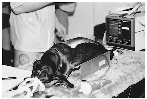
A five-year-old, male Dachshund is being prepared for emergency thoracolumbar spinal surgery. The owners found the dog dragging its hindlimbs about 4 hours earlier at 5 pm. The dog was noted to walk normally at 12 noon, but seemed wobbly at 2 pm. On your neurologic examination prior to any anesthetic agents, the cranial nerves and forelimbs were normal. There was some voluntary motor movement of both hindlimbs but the dog could not rise on its hindlimbs. A radiograph was taken with the dog under anesthesia.
| Question | Answer | Article | |
| Does this dog have deep pain, and how do you know? | Yes, because deep pain sensation is not lost until after voluntary motor movement is lost. |
[[|Link to Article]] | |
| How do you perform a deep pain response test, and what constitutes a positive deep pain response? | By applying a noxious stimulus, e.g. hemostats on the toes (to ‘crunch bone’). Conscious reaction to the stimulus, such as crying and turning to bite at the stimulus, constitutes a positive deep pain response. |
[[|Link to Article]] | |
| Where is the lesion based on this radiograph? (Assume that there are 13 ribs bilaterally.) | The T12–T13 intervertebral disc space. |
[[|Link to Article]] | |
| List six plain film radiographic signs of acute thoracolumbar disc extrusions. (Do not assume that all of these radiographic signs are present in this case.) |
|
[[|Link to Article]] | |
| What contrast radiographic study is used to localize the surgical lesion definitively? | Myelography. |
[[|Link to Article]] | |
| What are the three potential sources of pain in intervertebral disc disease? |
|
[[|Link to Article]] | |
| Why is paresis/paralysis less common with acute cervical intervertebral disc disease than with acute thoracolumbar disc disease? | Because there is a smaller ratio of spinal cord diameter to vertebral canal diameter. |
[[|Link to Article]] | |
| Give two reasons why corticosteroids must be used cautiously in acute intervertebral disc disease. |
|
[[|Link to Article]] | |
