Normal Intra-Oral Radiographic Anatomy - Small Animal
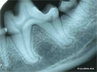
Teeth are divided into the crown, neck and root. The radiopaque structures of the tooth are mostly dentin covered by a thin layer of radiodense enamel (incompletely visualized on a radiograph) and cementum which is less dense than dentin and only really visible if it has undergone hyperplasia. The internal radiolucent area is the pulp cavity – it extends into the crown and down to the apex of the root. As the tooth matures, more dentin is laid down and the pulp size reduces with age. An immature tooth can be present in a mature animal where early trauma has caused pulp necrosis and thus stopped further development of the tooth. The tooth is attached to the boney socket by the periodontal ligament. The periodontal ligament space appears as a thin radiolucent line of uniform width between the root and bone. The cortical bone on the alveolar ridge appears as a radiopaque line of uniform thickness known as the lamina dura. The alveolar bone should extend to the cementoenamel junction.
The cervical area of the tooth, between the enamel of the crown and the alveolar bony margin, has neither enamel nor bone superimposed and is therefore less radiodense. This is referred to as “cervical burn-out” and should not be mistaken for caries or dental resorption. The bone of the alveolar margin should be relatively horizontal and positioned 1 to 2 mm apical to the cementoenamel junction. The interradicular marginal bone often has a slightly convex contour, filling the furcation area and closely approximating the contour of the furcation. In contrast, the interalveolar marginal bone (margin of septal bone) can have a horizontal, a slightly concave, or a slightly convex contour depending on the proximity of the adjacent roots and the particular region in the arch.
Nutrient Canals - The nutrient canals referred to here are those that contain the blood vessels and nerves that supply the teeth, interdental spaces and gingiva. In radiographs, these are seen as radiolucent lines of uniform width, which sometimes have radiodense borders. The most easily identified nutrient canal is the mandibular canal. Other nutrient canals, which may be seen, are the canal or groove that occupies the posterior superior alveolar artery and the anterior palatine (incisive) canal.
Mandibular Symphysis - A fibrous joint, present as a thin radiolucent irregular line on radiographs.
Foraminae - May sometimes be mistaken for periapical lesions. Important foramina to remember are: the anterior palatine (incisive) foramen, the infraorbital foramina and the mental foramina.
Mandibular Canal
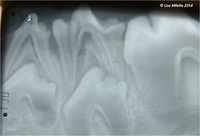
Deciduous Teeth – Thin narrow roots, may be resorbing, underlying tooth buds may be present.
Immature Permanent Teeth – Thin dentin, wide pulp, incomplete apex formation (open apex).
Mature Permanent Teeth – Thicker dentin, apex closed and thinner pulp.
| This article was written by Lisa Milella BVSc DipEVDC MRCVS. Date reviewed: 13 August 2014 |
| Endorsed by WALTHAM®, a leading authority in companion animal nutrition and wellbeing for over 50 years and the science institute for Mars Petcare. |

