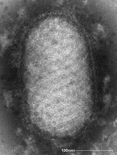
Poxviridae
Poxviruses are among the most easily recognized of all viruses, owing to the lesion by which they have gained their name. Once inside cell, they cause proliferation then lysis, giving way to a characteristic pock with a necrotic center. Poxviruses have risen to fame both for their ability to be eradicated (small pox) as well as their use in fighting other viruses (canarypox vaccines).
Content
- Small pox (variola)
- Pigeon pox
- Canary pox
- Camel pox
- Monkey pox
- Red squirrel parapox
- Red/Gray squirrel pox
Morphology
- Huge (up to 450nm), usually enveloped viruses, with a complex capsid symmetry
- Up to 30 different structural proteins
- Non-structural proteins:
- Viral epidermal growth factor, which stimulates cell growth causing the raised edge of pustule
- Viral tumor necrosis factor, which is non-functioning and acts as an anti-inflammatory by competing with TNF-alpha
- Viral IL-10, which reduces the Th-1 cell mediated response
Therapeutic Use
Recombinant Vaccines
- Poxviruses can be used as heat-stable vectors for vaccines against other viruses
- Grown in host cell lines or on the surface of chick chorioallantoic membranes in ovo (primordial ectoderm)
- This was first accomplished by the recombination of cowpox and variola (smallpox) in the creation of the smallpox vaccine (vaccinia)
- More recently, the French used this technique in the creation of the oral rabies vaccine used among the wild fox population:
- Recombinant virus inserts a plasmid encoding rabies gene in place of thymidine kinase gene
- Canarypox vaccines now exist for FeLV and Rabies
- Undergoes a single cycle of replication without producing infectious virus in mammals
Virulence and Pathogenesis
- Primary replication in abraded squamous epithelium
- Viremia followed by multiple epidermal infections
- Ballooning then necrosis (hydropic degeneration) of epidermal cells
- Concurrent proliferation or adjacent epidermis (GF driven), creating more cells for the virus to infect
- All three result in classical sequence of lesions:
- Papule (proliferation)
- Vesicle (fluid filled)
- Pustule (lesion breaks)
- Scab formation (healing begins)
- Pock center can succumb to secondary infection
- Resolution in 3-4 weeks
- Some poxviruses can spread to the upper respiratory tract or viscera, causing more serious pathology
Epidemiology
- Spread quickly in unhygienic circumstances
- Can survive for years in dust
Diagnosis
- Clinical signs
- Histology
- Electron microscopy
- PCR, IIF
Types and Subtypes
- 6 Genera, all of which produce pox lesions
- Subdivided based on external structure by EM
| Poxviridae Learning Resources | |
|---|---|
To reach the Vetstream content, please select |
Canis, Felis, Lapis or Equis |
Pages in category "Poxviridae"
The following 9 pages are in this category, out of 9 total.
