Conscious Oral Examination
Introduction
Oral examination of a conscious animal is limited to visual inspection and some digital palpation only. The aim of the conscious oral examination is to obtain a tentative diagnosis and help formulate a treatment plan. The owner must be made aware however that it is only under a general anaesthetic and full examination that the true extent of disease can be properly evaluated.
Approach
It is important to always have good lighting when examining the mouth (both in the conscious and anaesthetised patient).
Gentle technique is essential as some animals may have dental pain or discomfort. Examination involves assessing not only the oral cavity proper, but also palpation of:
- The face (facial bones and zygomatic arch)
- Muscles of mastication
- Temporomandibular joint
- Salivary glands (mandibular/sublingual; the parotids are usually only palpable if enlarged)
- Lymph nodes (mandibular, cervical chain)
- Eyes
Asymmetry of the head may be seen in congenital abnormalities, inflammatory diseases (cellulitis/abscess formation), neoplasia, dislocations, fractures, muscle wastage and nerve damage. Jaw movement should always be assessed to ensure that the mouth can open and close fully. The temporomandibular joints should be palpated during jaw movement to check for any crepitation or popping movements. To feel the temporomandibular joints, place three fingers over the joint (positioned rostral to the lateral ear canal, below the zygomatic arch) whilst a second person gently opens the mouth. A unilateral exophthalmia may result from a retrobulbar or orbital abscess. It is also useful to apply gentle pressure to the eye to assess pressure if a lesion in this area is suspected.
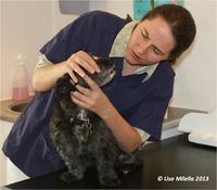
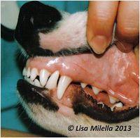
The mouth is first examined by gently holding the jaws closed and retracting the lips (do not pull on the fur to retract lips) to look at the soft tissues and buccal aspects of the teeth. Check the lip for clefts, lacerations, inflammation, ulcerations, or swellings before lifting the lip to examine the soft tissues and teeth. Apart from colour and texture of the mucous membranes, look for evidence of a potential bleeding problem (petechiation, purpura, ecchymoses). In addition, look for vesicle formation and ulceration, which could indicate a vesiculo-bullous disorder, e.g. pemphigus, pemphigoid. It is useful to count the teeth to ensure that a tooth is not absent without reason. Obvious dental pathology (tooth fracture, gingival recession, advanced furcation exposure) can also be identified. It is useful to compare left and right sides of the mouth – animals in pain may be chewing on one side only, resulting in less plaque and calculus on the side of the mouth that is being used.
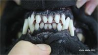
With the lips gently retracted and the mouth closed, this is the optimal time to evaluate occlusion.
6-point checklist for dental occlusion:
- Head symmetry
- Incisor relationship (the lower incisors usually occlude on the palatal aspect of the upper incisors in a mesocephalic skull type). This is referred to as a scissor bite.
- Canine occlusion (the lower canine occludes equidistant between the upper canine and the third incisor. The cusp should be visible on the buccal aspect of the gingiva and should not be touching any soft tissue)
- Premolar alignment (the premolars interdigitate in a pinking shear effect. The main cusp of the lower premolar should bite between the two upper corresponding premolars)
- Distal premolar/molar occlusion (the mandibular premolars and molars occlude on the palatal aspect of the uppers)
- Individual teeth positioning
It is not unusual for dogs and cats to have abnormal occlusion. When assessing whether treatment is required or not, look for any tooth contacting soft tissue and causing trauma, or any tooth-on-tooth contact resulting in trauma.
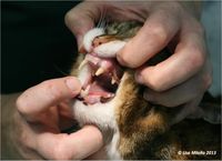
Finally, the animal is encouraged to open its mouth. One method, useful for both dogs and cats, is to approach the animal from the side, one hand is placed over the muzzle and the lips are gently pressed into the oral cavity, while tilting the head slightly upwards. A finger from the other hand is placed on the lower incisors and gentle pressure is exerted. Do not use the fur under the mandible to try to pull the jaw down. Most animals allow at least a cursory inspection of the oral cavity once the jaws have been opened. The mucous membranes of the hard palate and the oropharynx (soft palate, palatoglossal arch, tonsillary crypts, tonsils and fauces) can be assessed. The occlusal surfaces of the molar teeth as well as the lingual and palatal surfaces of the teeth can be briefly evaluated.
Some animals may be uncomfortable and have painful areas in the mouth, limiting conscious examination. Difficulty examining the mouth may be an indication to sedate or anaesthetize the patient to examine the mouth fully.
Limitations of Conscious Examination
- Pain may make the animal less co-operative
- Not all surfaces of the tooth are easily visible during conscious examination
- The periodontal status cannot be assessed in a conscious patient as the area below the crown/under the gum margin cannot be assessed.
- Sometimes pathology can be identified but the full extent of the pathology cannot be assessed until further diagnostic tests, such as intra-oral radiographs have been taken. Eg. Feline odontoclastic resorptive lesions
- Only a tentative diagnosis and treatment plan can be established, hence treatments involved and cost cannot be accurately estimated at this time. This is one of the most important aspects of client communication to avoid conflict after treatment.
Download a Dental Decision Guide:
| This article was written by Lisa Milella BVSc DipEVDC MRCVS. Date reviewed: 12 September 2014 |
| Endorsed by WALTHAM®, a leading authority in companion animal nutrition and wellbeing for over 50 years and the science institute for Mars Petcare. |
Error in widget FBRecommend: unable to write file /var/www/wikivet.net/extensions/Widgets/compiled_templates/wrt69a03e7ed0fd15_82690248 Error in widget google+: unable to write file /var/www/wikivet.net/extensions/Widgets/compiled_templates/wrt69a03e7ed63da7_09534440 Error in widget TwitterTweet: unable to write file /var/www/wikivet.net/extensions/Widgets/compiled_templates/wrt69a03e7edd1da0_61143994
|
| WikiVet® Introduction - Help WikiVet - Report a Problem |


