Large Intestine - Anatomy & Physiology
Introduction
The large intestine extends from the ileum of the small intestine to the anus. Water is absorbed from the ingesta and faeces are stored prior to defeacation. Every species has a large microbial population living in the large intestine, which is of particular importance to the hindgut fermenters. For this reason, hindgut fermenters have a more complex large intestine with highly specialised regions for fermentation.
The large intestine can be divided into:
Structure
Function
Vasculature
- The cranial mesenteric artery supplies the caecum, ascending and part of the transverse colon.
- The caudal mersenteric artery supplies the rest of the transverse colon, the descending colon and the rectum.
Innervation
- Like the small intestine, the large intestine recieves sympathetic and parasympathetic innervation.
- Neurones interact with the myenteric plexus to affect contractility, and with the submucosal plexus to affect secretions.
- The sympathetic have coeliac, cranial mesenteric and caudal mesenteric ganglia.
- As the sympathetic fibres leave the ganglia, they surround their respective artery.
- Parasympathetic innervation stimulates peristalsis.
Lymphatics
- Lymphatic nodules are present in the mucosa.
Histology
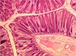
- The mucosa of the large intestine is smooth; there are no villi.
- Mucosal glands are much longer and straighter.
- The number of goblet cells in the mucosa is increased compared to the small intestine.
- Mucus is very important for lubrication of the ingesta as it passes through the intestine, particularly as more water is absorbed from the lumen making it drier.
- There are numerous scattered lymph nodules.
- The number of lymph nodules increases compared to the small intestine.
- Taenia may be present.
- These are concentrations of the longitudinal muscle layer into long bands.
- When the taenia contract, they cause shortening of the large intestine, which produces saccualtions, or haustra.
- Many glands are present in the mucosa and skin of the anal region.
Species Differences
Carnivore
- The dog and cat posses two anal sacs. In the dog, these are the size of a hazlenut.
- They are located ventrolaterally between the internal and external anal sphincters.
- The fundus of the sac secretes a potent smelling fluid that drains through a single duct to an opening near the anocutaneous juncntion.
- The anal sacs get compressed during defecation, which causes the fluid to be expressed. The scent of the fluid is thought to act as a territorial marker.
Ruminant
Horse
Pig
- Taenia present
