Small Animal Emergency and Critical Care Medicine: Self-Assessment Color Review, Second Edition, Q&A 07
Jump to navigation
Jump to search

| This question was provided by CRC Press. See more case-based flashcards |

|
Student tip: This case is a lesson in how to infer diagnosis from biochemical results. |
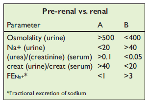
A 3-year-old female Daschund presents for vomiting eight times in the last 24 hours. T = 39.4°C (103°F); HR = 180 bpm; RR = panting; CRT = >3sec; MM gray, dry; femoral pulses weak; perfusion - early decompensatory shock; 8% dehydrated by skin turgor, dry MM/corneas. Biochemistry laboratory abnormalities: BUN = 28.5 mmol/l (80 mg/dl); creatinine = 177 μmol/l (2.0 mg/dl); K+ = 3.0 mEq/l; lactate = 3.4 mmol/l (30.6 mg/dl); pH = 7.31; HCO3 = 15 mEq/l; PCO2 = 30 mmHg.
| Question | Answer | Article | |
| It is often difficult to differentiate pre-renal azotemia from renal azotemia. Given these tests and results (see table above), which column is pre-renal and which is renal? | Column A is pre-renal; column B is renal.
|
Link to Article | |
| List markers of kidney disease found on blood tests and on urinalysis. | Blood: elevated BUN; elevated creatinine; hyperphosphatemia; hyperkalemia; hypokalemia; metabolic acidosis; hypoalbuminemia. Urine: isosthenuria; proteinuria; cylinduria; renal hematuria; inappropriate urine pH; glycosuria.
|
Link to Article | |
| List four proposed mechanisms for oliguria in AKI. | Vascular theory: vasoconstriction of pre- and post-glomerular arterioles making renal glomerular capillary pressure inadequate for filtration. Tubular swelling: damage to tubular cells causes swelling and dysfunction. Tubular obstruction: damaged tubular cells exfoliate, creating a cast that obstructs the urine outflow from that tubule. Tubular back leak: fluid and molecules from within the renal tubule back leak into the peri-tubular capillary.
|
Link to Article | |
| Gram-negative rods were seen in the abdominal fluid in this dog, found sensitive only to an aminoglycoside antibiotic. Describe how to monitor this dog for early evidence of nephrotoxicity caused by the aminoglycoside. | Nephrotoxicity is due to altered phospholipid metabolism as the drug accumulates in the proximal tubular cells, made worse by poor perfusion and dehydration. Monitor: urine sediment for increasing presence of casts; glycosuria with normal blood glucose; increasing BUN and/or creatinine (occurs later than urine changes); BP, urine output; physical perfusion and hydration parameters; body weight; and occasionally serum drug concentrations.
|
[[ Replace text with name and subsection of relevant WikiVet page if in existence eg. Feather - Anatomy & Physiology#Structure & Function |Link to Article]] | |
To purchase the full text with your 20% off discount, go to the CRC Press Veterinary website and use code VET18.
