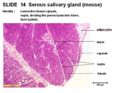
Serous Salivary Gland - Anatomy & Physiology
Jump to navigation
Jump to search
Overview
The serous salivary gland has a connective tissue capsule and septa dividing the parenchyma into lobes. There is a duct system. Interlobular ducts run in the tissue septum lined by cuboidal to columnar epithelium.
Intralobular ducts run within the lobules. Striated intralobular ducts are lined with cuboidal epithelium. Intercalated intralobular ducts are lined with low cuboidal to simple squamous epithelium. Serous acini secrete a watery solution rich in proteins with spherical nuclei. Cells are pyramidal, cuboidal or crescent shaped.
| Serous Salivary Gland - Anatomy & Physiology Learning Resources | |
|---|---|
To reach the Vetstream content, please select |
Canis, Felis, Lapis or Equis |
 Test your knowledge using flashcard type questions |
Salivary Glands Anatomy & Physiology Flashcards |
 Selection of relevant PowerPoint tutorials |
Oral Cavity Histology, see part 2 for salivary glands |
Webinars
Failed to load RSS feed from https://www.thewebinarvet.com/dentistry/webinars/feed: Error parsing XML for RSS
