Pleural Cavity and Membranes - Anatomy & Physiology
Jump to navigation
Jump to search
|
|
Introduction
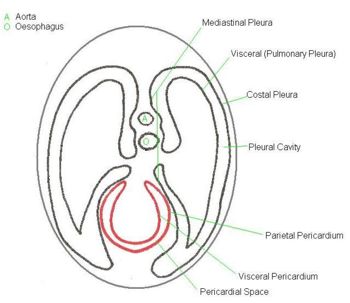
The surface of the inner wall of all of the body cavities are lined by a serous membrane which consists of a single layer of flat epithelium with a thin underlying propria (connective tissue). Within the thoracic cavity, this is known as the Pleura.
Structure of the Pleural Membranes
- Each lung is placed within a separatte layer of membrane, thus there are two pleural sacs.
- The space between the two sacs is known as the Mediastinum.
