Nasal Cavity - Anatomy & Physiology
|
|
Introduction
The respiratory tract begins with the nose which includes the external nose, internal nasal cavities and paranasal sinuses. As well as being vital for transport of gases to the lower respiratory tract, the nose is also the site for one of the special senses - Olfaction.
Structure
- The nose consists of the external nares with nasal cartilages, the nasal cavity (including the nasal meatus and conchae), and the paranasal sinuses.
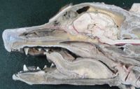
- The nasal cavity is essentially a tube with a wall established by several bones of the skull. The borders of the nasal cavity are as follows:
- Caudal: cribrifrom plate of the ethmoid bone
- Ventral: continuous with the nasopharynx
- Dorsal: the maxilla and the palatine processes of the incisive bone
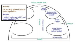
- Rostrally, the median septum is a continuation of the ethmoid bone. The median septum is made up of hyaline cartilage, and divides the nasal cavity into left and right halves.
- The nasal cavity is occupied to a large extent by Nasal conchae. These are turbinate bones which project into the nasal cavity with the purpose of supporting the olfactory mucus membranes and increasing the respiratory surface area, creating turbulence within the passing air. This helps to filtrate and warm or cool the air that passes through.
- The Conchae are cartilagenous or ossified scrolls which arise from the ethmoid bone. They are covered with mucous membrane, under which is a layer of anastomosing blood vessels.
The nasal conchae are more complex in animals with a better sense of smell, as they increase the surface area of the Olfactory region, further.
- There are dorsal and ventral conchae, the dorsal concha originating from the ethmoid bone and attaching to the maxilla, and the vental conchae originating from the maxilla and extending further into the nasal cavity.
- The conchae divide the nasal cavity into nasal ducts or meatuses, which branch out from a common nasal meatus which is adjacent to the nasal septum. There are three nasal meatuses which branch from the common nasal meatus: dorsal, middle and ventral:
- Dorsal nasal meatus: the passage between the roof of the nasal cavity and the dorsal nasal concha
- Middle nasal meatus: between the dorsal and ventral conchae, and communicates with the paranasal sinuses.
- Ventral nasal meatus: the main pathway for airflow leading to the pharynx, and is positioned between the ventral nasal concha and the floor of the nasal cavity.
- Common nasal meatus: the longitudinal space on either side of the nasal septum.
- The Paranasal Sinuses are extensions of the nasal cavity.
Function
- In addition to Olfaction, the function of the nasal cavity is to modify the incoming air before is is transported further down the respiratory tract.
- Air is warmed as it passes over the highly vascularised mucosal surfaces of the conchae, humidified by the evaporation from nasal secretion and cleaned as it contacts the secretion from mucus glands within the nasal cavity. The mucus secreted from the glands traps particles and cilia transport them down to the pharynx for swallowing, this process is known as the Mucociliary Escalator.
- The nasal cavity offers further protection via the Sneezing reflex .
Species Differences
- The nasal cavity in the sheep is highly vascularised, with any damage to the epithelium resulting in severe haemorrhage.
- Cattle have a smaller nasal cavity compared to the horse.
- There are many variations to the entire Avian respiratory tract.
- The Respiratory Systems of non-Homeotherms]] are also very different to that of mammals.
Links
References
- Dyce, K.M., Sack, W.O. and Wensing, C.J.G. (2002) Textbook of Veterinary Anatomy. 3rd ed. Philadelphia: Saunders.

