Adrenal Glands - Anatomy & Physiology
|
|
Adrenal Glands
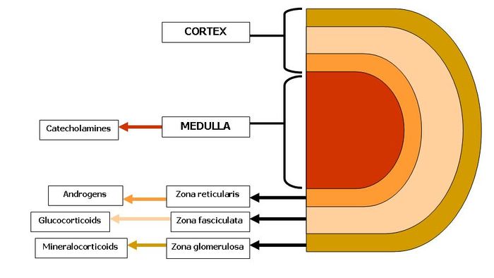
The adrenal glands are paired bodies lying cranial to the kidneys within the retroperitoneal space. The glands consist of two layers; the cortex and medulla.
The adrenal cortex is red - light brown in colour and is comprised of three zones. These zones all produce hormones derived from cholesterol which is abundant in the cells. From outer to inner the layers are zona glomerulosa, zona fasciculata and zona reticularis. The adrenal cortex represents 80-90% of the adrenal gland.
The adrenal medulla is primarily involved in the production of catecholamines; epinephrine and norepinephrine. In fetal life the adrenal medulla plays a role in the autonomic nervous system. The medulla acts as a sympathetic ganglion with the postganglionic cells lacking axons. Through sympathetic preganglionic fiber stimulation the medullary cells secrete catecholamines. The adrenal medulla represents only 10-20% of the adrenal gland.
Embryological Origin
The adrenal glands develop from two separate embryological tissues; the neural crest ectoderm and the intermediate mesoderm. The adrenal cortex develops from the intermediate mesoderm. The medulla originates from neural crest cells migrating from sympathetic ganglion. Mesodermal cells then surround the medulla.
The fetal cortex develops in the centre with the permanent cortex surrounding it. By 4 months of age the adrenal gland is fully developed.
Anatomy
Vascular Supply
Function
Histology of the Adrenal Glands
<gallery> Image:Histology of the Adrenal Glands..jpg|Histological section of the Adrenal Gland Image:Histology of the Adrenal Glands showing zones..jpg|Histological section of the Adrenal Gland Cortical zones Image:Histology of the Adrenal Glands Medulla..jpg|Histological section of the Adrenal Gland showing Medulla and Zona Reticularis <gallery>
