File:Giardia cysts.jpg
Giardia_cysts.jpg (369 × 235 pixels, file size: 9 KB, MIME type: image/jpeg)
H.D.A. Lindquist, U.S. EPA
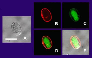
The same Giardia lamblia (intestinalis) cyst as imaged at different instrument settings by confocal microscopy. (A) is the cyst imaged by transmission (differential interference contrast), only. (B) is the cyst wall selectively imaged through use of fluorescent-labelled (TRITC) antibody that is cyst wall specific. (C) is the cyst imaged through use of carboxy fluorescein diacetate, a viability stain. (D) is a composite image of (B) and (C). (E) is a composite image of (A), (B), and (C). Bar = 10 microns.
Source: Gerbil stool.
Method: Confocal Microscopy
File history
Click on a date/time to view the file as it appeared at that time.
| Date/Time | Thumbnail | Dimensions | User | Comment | |
|---|---|---|---|---|---|
| current | 18:08, 23 November 2008 | | 369 × 235 (9 KB) | Nabrown (talk | contribs) | H.D.A. Lindquist, U.S. EPA The same Giardia lamblia (intestinalis) cyst as imaged at different instrument settings by confocal microscopy. (A) is the cyst imaged by transmission (differential interference contrast), only. (B) is the cyst wall selectively |
You cannot overwrite this file.
File usage
The following file is a duplicate of this file (more details):
- File:Giardia.jpg from Wikimedia Commons
There are no pages that use this file.