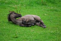
Category:Colic in Horses
Colic in horses is defined as abdominal pain, and can be caused by a wide variety of conditions. Many of these conditions are life threatening, and therefore it is essential to diagnose and treat cases of colic as quickly as possible. The most common causes of colic are gastrointestinal conditions, although it can also be caused by other abdominal conditions. In the latter case, it is often called false colic. Treatment of colic is largely dependent upon identifying the underlying reason for the pain, and treating this cause appropriately. Most commonly this is done medically, but in a small percentage of cases, surgical intervention is needed. Among domesticated horses, colic is a major cause of premature death. The incidence of colic in the general horse population has been estimated between 10 and 20 percent on an annual basis. It is important that any person who owns or works with horses be able to recognize the signs of colic, so that a veterinarian may be called promptly, before the horse's condition deteriorates.
Surgical Conditions
Small Intestine
Impaction
Association with ascarid infection[1]
Intussusception
This is a condition in which one part of the intestine "telescopes" inside another. Usually this obstructs the blood flow to the inner part, and so forms a strangulating obstruction. Intussusception can occur within the small intestine, and also between small intestine and caecum (ileo-caecal intussusception). The latter is predisposed by Anoplocephala perfoliata tapeworm infection. When working up an acute abdominal case, it must be borne in mind that this form of colic is serious and necessitates surgery, however, peritoneal fluid changes will not usually be seen, as will often be found in a surgical colic. This is because the strangulated portion of gut (the inside of the "telescope"), is contained within an intact piece of intestine, so leaking fluid and protein is contained from the peritoneal cavity.
Herniation/entrapment
- Inguinal canal
- Umbilical hernia
- Epiploic foramen
- Mesenteric rents/tears
- Diaphragmatic hernia
- Mesodiverticular bands
- Gastrosplenic ligament
Pedunculated lipoma
Volvulus (nodosus)
Rotation of mesenteric root
Caecum
Caeco-caecal intussusception
Torsion
Impaction
Large Colon
Torsion
Left dorsal displacement
Right dorsal displacement
Sand impaction
Enterolith
Small Colon
Faecolith
Enterolith
Strangulating lipoma
Any location
Foreign body
False colic
Signs of colic may be caused by abdominal pain not associated with the gastro-intestinal tract, for example, pain associated with uterine or testicular torsion, or originating from the kidneys, liver, ovaries, spleen, pleuritis, or pleuropneumonia. Other diseases which sometimes cause symptoms which appear similar to colic include laminitis and exertional rhabdomyolysis.
Clinical signs
- Pawing and/or scraping
- Stretching
- Frequent attempts to urinate
- Flank watching
- Pacing
- Repeated flehmen response
- Repeated lying down and rising
- Rolling
- Groaning
- Bruxism
- Excess salivation
- Inappetance
- Decreased faecal output
- Increased pulse rate
- Dark mucous membranes
Medical Treatment
Surgical Treatment
Post-surgical Management
Prevention
Epidemiology
Incidence
Colic occurs relatively frequently in horses, with an incidence estimated at 0.1-0.2 episodes per horse-year. In context, this would mean an average holding of 100 horses could reasonably expect to see 10-20 cases every year.
Classification
Approximately 90% of colic episodes can be succesfully managed using medical treatments, with the remainder requiring surgery. Assuming surgical and medical cases of colic are accurately distinguished, survival rates of 95% and 80% are considered normal for medical and surgical colic, respectively.
Post-operative Survival
Studies have shown that there is an increased risk of death with certain factors:
- Abnormal Packed Cell Volume (PCV) on presentation
- Increased length of intestine resected
- Increased duration of surgery
- Elevated peripheral lactate
- Elevated peritoneal fluid lactate
See Also
Further Reading
References
- ↑ Cribb NC, Cote NM, Bouré LP, Peregrine AS. (2006). Acute small intestinal obstruction associated with Parascaris equorum infection in young horses: 25 cases (1985-2004).. New Zealand Veterinary Journal
Subcategories
This category has the following 9 subcategories, out of 9 total.
A
B
C
D
E
F
I
M
S
Pages in category "Colic in Horses"
The following 5 pages are in this category, out of 5 total.
