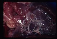
Category:Cardiovascular System - Developmental Pathology
Introduction
Congenital abnormalities occur relatively commonly in young (approximately 1% all human births). May produce dramatic clinical signs or signs may be vague, such as failure to thrive. Clinical signs may become more apparent as the animal grows and activity levels increase.
The aetiology of congenital defects of the heart and great vessels are mostly unknown. Possible contributing factors include:
- Genetic; E.g. Mutation in the ova or sperm or mutation in the zygote.
- Environmental; Includes infection (usually viral), chemical, nutritional and physical factors.
Understanding the normal development of the heart and great vessels allows an understanding of the morphology and pathology of the congenital abnormalities seen in mammals.
Normal Development
The heart develops from a simple endothelial tube which acquires a mesodermal layer (forms myocardium and epicardium). As the heart grows a flexure forms so that the original cranial portion becomes the truncus arteriosus and the ventricles and the oringinal caudal part of the tube will form the sinus venosus and the atria.
The atria are separated by a growth from the dorsal wall, the septum primum and ventral wall, the septum secundum. The ventricles are separated by a septum growing from the apex.
Congenital abnormalities can be broadly classified into 3 groups:
- Abnormalities of chamber development forming shunts.
- Abnormalities of great vessels forming shunts or anomalies.
- Abnormalities of valve formation resulting in dysplasias.
Chamber Development
Atrial Septal Defect
Persistent Foramen Ovale, allowing blood flow from left to right atria. Produces volume overload of right ventricle and subsequent raise in central venous pressure and venous congestion.
Incidence:
- Rare in cats and dogs, increased incidence in Old English Sheepdogs and Boxers.
- More common in cattle, often due to failure of growth of septum secundum.
Clinical Signs:
- If small defect no clinical signs.
- If large defect see exercise intolerance, syncope and dyspnoea.
Diagnosis:
- Enlarged right ventricle and atrium on radiology.
- Can visualise on echocardiography.
Treatment:
Usually no management needed.
Ventricular Septal Defect
Often associated with other conditions E.g. Tetralogy of Fallot.
Usually occur high in the membranous region of the interventricular septum. Blood flows from left to right ventricle during systole resulting in right ventricular overload, increased central venous pressure and pulmonary overcirculation.
Eisenmenger's Syndrome: chronic pulmonary overcirculation results in pulmonary hypertension and increased right-sided pressure. This facilitates right to left shunting and reduced lung perfusion, leading on to cyanosis.
Incidence:
- Not common in dogs but increased incidence in WHWTs and Cocker Spaniels.
- More common in cats and cattle.
Clinical Signs:
- Depend upon size of defect, may include; exercise intolerance, venous congestion etc.
- Harsh holosystolic murmur. Murmur grade correlates inversely with the defect size.
Diagnosis:
- Left and right ventricular hypertrophy, often generalised cardiomegaly on radiology and ECG.
- Pulmonary overcirculation on radiology.
- Left ventricle hyperkinetic on echocardiography and often the defect may be visualised. Doppler echocardiography may be useful.
- Mitral regurgitation is common due to a change in size of the left ventricle and consequent alterations to the Mitral valve.
Treatment:
- Small defects require no treatment.
- Pulmonary Banding protects the pulmonary circulation by restricting its blood flow.
- Symptomatic therapy for left heart failure E.g ACE inhibitors.
- Prognosis variable with defect size.
Patent Ductus Arteriosus
Ductus Arteriosus shunts blood from the Pulmonary Artery to the Aorta in the foetus to bypass the lungs. Usually closes shortly after birth due to smooth muscle contraction in repsonse to increased arterial oxygen tension. When patent after birth, blood flows along the pressure gradient, left to right, leading to pulmonary hypertension. If the hypertension is significant, as with larger defects, right sided pressure may increase enough to allow reversal of flow through the PDA and will result in cyanosis. Reverse shunting is rare.
Incidence:
- Uncommon in cats.
- More common in dogs with distinct female predisposition.
- Predisposed breeds include; CKCS, Border Collie, GSD, Poodles.
- Research into PDAs in dogs has revealed a genetic component influencing the maturation of collagen.
- May be a normal finding in lambs and piglets up to 2 weeks of age.
Clinical Signs:
- Continuous machinery murmur at left heart base.
- Water-Hammer pulse (rapid diastolic decline).
- Variable other signs depending on size of defect E.g. syncope, cyanosis, exercise intolerance etc.
- May progress to left sided heart failure and so pulmonary oedema.
Diagnosis:
- Left atrial and left ventricular enlargement radiologically and on ECG due to volume overload.
- Pulmonary overcirculation (enlargement of pulmonary arteries and veins) on radiology.
- DV radiographs show a three knuckled appearance; enlargement of:
- Aortic arch.
- Pulmonary artery.
- Left auricular appendage.
- PDA difficult to image on echocardiography but Doppler imaging may be useful.
Treatment:
- Surgical ligation for left to right shunts. Needs stabilisation first.
- Right to left shunts are not operable.
- Untreated dogs face 60-70% mortality during their first year of life.
Great Vessels
Vascular Ring Anomaly
Most commonly found in dogs and cats. Acyanotic; clinical signs due to constriction of the oesophagus:
- Regurgiation of food, usually noticed at weaning.
- Aspiration pneumonia.
Persistent Right Aortic Arch
Most common vascular ring anomaly, increased incidence in GSDs, and Irish setters. vascular ring forms between the ductus arteriosus/ligamentum arteriosum and the persistent right aorta. Megaoesophagus is seen cranial to the constriction.
Other causes include:
Double aortic arch
Anomalous subclavian arteries
Arise form the aortic arch rather than the brachiocephalic artery.
Diagnosis:
- Ventral deviation of the trachea and dilation of the oesophagus on radiology.
- Barium swallow definitive.
Treatment:
- Surgical resection possible.
- Dietary management usually sufficient to control regurgitation.
Tetralogy of Fallot
- Pulmonic stenosis
- Right ventricular hypertrophy
- High VSD
- Overriding aorta
Commonly reported in certain breeds, E.g Keeshond, Schnauzer, Poodles and terriers.
Clinical Signs:
- Cyanotic; see severe exercise intolerance.
- Systolic murmur due to pulmonic stenosis +/- precordial thrill.
- Polycythemia due to chronic hypoxia.
Diagnosis:
- Right ventricular enlargement on radiology and ECG.
- Post stenotic dilatation of pulmonary artery on radiology and pulmonary undercirculation due to right to left shunting.
- Echocardiography may show right ventricular hypertrophy, paradoxical motion of interventricular septum (pressure RV>LV), visulaisation of VSD and overriding aorta.
- Doppler useful to image pulmonic stenosis and VSD.
Valve Development
Aortic Stenosis
Obstructs blood flow from the left ventricle to the aorta. Stenosis may be:
- Subvalvular (most common in dog and cat).
- Valvular.
- Supravalvular.
Subvalvular stenosis results in a jet of blood hitting the aortic valve, leading to damage and increased risk of developing endocarditis.
Incidence:
- Increased prevalence in Boxers and Golden retrievers.
- Autosomal dominant in Newfoundlands.
Clinical Signs:
- Vary from asymptomatic to degrees of exercise intolerance, syncope and even sudden death.
- Harsh systolic murmur at left heart base.
Grade correlates with grade of murmur. High grade murmurs also have a precordial thrill.
- Murmur may have a diastolic component due to aortic valve damage and insufficiency.
- Weak femoral pulse.
Diagnosis:
- Left ventricular enlargement on radiology and ECG.
- Post-stenotic dilatation of the aorta may be visible on radiographs.
- Echogenic fibrous ring may be seen on echocardiography. Again, Doppler is useful to detect flow rates.
Treatment:
- Balloon valvuloplasty; often unsuccessful.
- Mild cases need no management.
- Medical management; Beta-blockers to decrease myocardial oxygen consumption.
- Prognosis guarded, death due to arrythmias in abnormal myocardium.
Pulmonic Stenosis
Obstructs blood flowing form the right ventricle to the pulmonary artery. As with aortic stenosis may be subvalvular, valvular or supravalvular. Valvular stenosis is the most common form in the dog.
Incidence:
- Increased incidence in English bulldogs, Cocker spaniels, Chihuahuas.
- Autosomal dominant in Beagles.
Clinical Signs:
- Asymptomatic if mild or moderate.
- If severe; exercise intolerance, stunting, syncope, right sided heart failure E.g. ascites.
- Harsh systolic murmur at left heart base.
- May hear splitting of the second heart sound due to delayed closure of the pulmonic valve.
- Femoral pulse quality good but jugulars distended.
Diagnosis:
- Right ventricular enlargement on radiology, along with post-stenotic dilatation of the pulmonary artery and pulmonary undercirculation.
- Abnormal valve can be imaged using echocardiography.
- Severity of stenosis can be assessed using Doppler to calculate flow rates.
Treatment:
- Balloon valvuloplasty is treatment of choice.
Mitral Dysplasia
Often associated with mitral regurgitation and left atrial volume overload. Usually progresses to left sided heart failure.
Incidence:
- Most common congenital defect in cats.
- Also reported in pure breed dogs E.g GSD, Great Danes.
Clinical Signs:
- Often murmur is the only clinical sign; pansystolic with increased intensity over the mitral valve area.
- May also see exercise intolerance, dyspnoea and coughing.
Diagnosis:
- Left atrial enlargement on radiology and ECG.
- Doppler echocardiography can dtect abnormal flow.
Treatment:
- Prognosis poor, medically manage left heart failure with ACE-inhibitors etc.
Tricuspid Dysplasia
Tricuspid regurgitation results in volume overload of the right atrium and may progress to right heart failure.
Ebstein's Anomaly: Atrioventricular ring is displaced ventrally into the right ventricle so that part of the wall of the right atrium is made up of ventricular wall.
Incidence:
- Second most common congenital anomaly seen in cats.
- Also reported in large breeds of dog including Weimeraner.
Clinical Signs:
- Often the murmur is the only clinical sign; low grade and pansystolic with increased intensity over the tricuspid area.
- May also see right sided failure, signs include; ascites, hepatomegaly and splenomegaly.
Diagnosis:
- Right ventricular enlargement on radiology and ECG.
Miscellaneous
Peritoneo-pericardial Diaphragmatic Hernia
Represents an embryological malformation of the ventral midline with communication to varying degress of the pericardial sac and the peritoneal cavity. Often associated with other malformations for example sternal deformities and ventricular septal defects. Usually results in gastrointestinal or respiratory signs. Visible radiologically as a round cardiac silhouette which merges with the diaphragm. The trachea will be displaced dorsally. Intestines may be visible on radiographs. Repaired surgically.
Ectopia Cordis
The heart is normal in anatomy but situated in an abnormal location within the body. Cattle and pigs show the highest incidence. The heart may be found in the cervical region or the abdomen.
Endocardial fibro-elastosis
Seen in Burmese kittens. Collagen and elastin are deposited in the endocardium, limiting its function. Animals are often found dead at 2-3 months of age.
Pages in category "Cardiovascular System - Developmental Pathology"
The following 14 pages are in this category, out of 14 total.
