Johne's Disease
Jump to navigation
Jump to search
| Also known as: | Midges |
| The most important veterinary species | Culicoides |
Paratuberculosis/ Johne's Disease is caused by Mycobacterium avium subsp. paratuberculosis, commonly seen in cattle, but known to affect all ruminants. Common signs include enteritis and diarrhoea.
- May be seen in zoo ruminants and goat herds.
- Particularly prevalent in Channel Island breeds.
- Is now also becoming a problem in Limousin breeds.
- Produces a chronic proliferative enteritis.
- Is usually fatal, since the disease cannot be got rid of.
- Animals may sometimes be carriers without showing clinical signs.
- Once disease is present in a herd, it is very difficult to get rid of it.
- Mycobacterium is excreted in urine and milk as well as in the faeces.
Clinical
- Clinical signs develop in older cows after calving i.e. 3 to 4 years of age.
- BUT animals are infected as calves less than 6 months old
- The disease develops very slowly.
- Clinical signs include:
- Ongoing, chronic profuse diarrhoea.
- Paint like consistency.
- Hindquarters and tail-caked with faeces
- Faeces also splattered on walls.
- Animal gradually fades away and dies over the course of months.
- Ongoing, chronic profuse diarrhoea.
Pathogenesis
- Organisms get in through the M-cells of Peyer's patches.
- Mycobacteria invade macrophages and cause a granulomatous inflammatory response.
- Death results from:
- Damage to the mucosa.
- Nutrients cannot be absorbed.
- Inflammatory loss of protein
- I.e. a protein losing enteropathy (hypoalbuminaemia).
- Damage to the mucosa.
Pathology
Gross
- Quite typical
- Cows appear very emaciated.
- Depends on how long the disease has been there.
- Not very much to see!
- Fat is pale and oedematous, and there is not much of it.
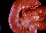
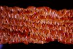
- Signs are confined to the terminal small intestine (especially the ileum) but are characteristic.
- Diffusely thickened mucosa
- Transverse, corrugated ruggae with reddened crests.
- Cannot extend the gut to remove these (i.e. they are permanent ruggae).
- Velvety mucosal surface.
- Mucosa may take on a 'corn-on-the-cob' appearance in advanced cases.
- Transverse, corrugated ruggae with reddened crests.
- Serosal oedema.
- Distended lymphatics.
- Diffusely thickened mucosa
- Enlarged mesenteric lymph nodes.
- Changes are milder in sheep and goats.
- Often missed.
- May produce small areas of necrosis not usually seen in cattle.
- Sheep may get a pigmented form.
Histologically
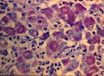
- Many large macrophages (epithelioid macrophages) in mucosa, submucosa and lymph nodes.
- Mesenteric lymph nodes are pale and enlarged (though not necrotic).
- The lamina propria is infiltrated by sheets of macrophages with some lymphocytes.
- Acid-fast bacteria are found in the macrophages and giant cells.
- Detected by Ziehl-Neelson stain.
- Bacteria act like foreign body producing a type IV hypersensitivity reaction.
- Sheep have two different forms.
- Paucibacillary
- Many T cells
- Few bacilli
- Multibacillary
- Many macrophages
- Many bacilli in macrophages
- Few lymphocytes
- Paucibacillary


