Canine Cyclic Haematopoiesis
| This article is still under construction. |
| Also known as: | Grey Collie Syndrome Cyclic Neutropaenia |
Description
Cyclic haematopoiesis is a disease inherited in an autosomal recessive manner. It is associated with an insertion mutation in the gene AP3B1 and it results in defective production of blood cells by bone marrow stem cells. The disease has only been described in grey collies which are homozygous for the genetic mutation; heterozygous animals do not show clinical signs.
From around 7 weeks of age, affected animals begin to show cyclic waves of neutropenia and of other cell type reductions. The reductions in other cell types (including platelets, eosinophils and red blood cells) do not occur in synchrony with each other or the neutropenia and bouts of reduction are interspersed with periods of marked blood cell overproduction. The periods of neutropenia last for approximately 2-4 days and are associated with severe systemic infections, including pneumonia, oronasal infection, bacterial enteritis, septic arthritis and sepsis.
During the periods when cell numbers are increased, it is thought that cytokines drive the production and deposition of amyloid proteins in various tissues, including the kidney, liver, spleen and pancreas. Almost all affected animals develop this further disease and it ultimately results in organ failure.
Affected animals rarely survive to sexual maturity and the disease is transmitted by heterozygous carriers.
Signalment
The disease has only been described in grey collies which are homozygous for the genetic mutation.
Diagnosis
Clinical Signs
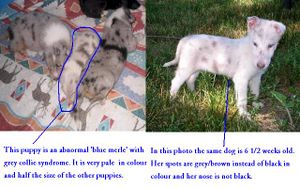
Affected animals have a diluted grey coat colour and are often smaller than their littermates. The majority of other clinical signs relate to the reductions in various types of cell, including:
- Pyrexic, lethargy and depression due to infection which develops rapidly in neutropenic animals.
- Signs of specific infections, including dyspnoea, diarrhoea, oral, nasal and ocular discharge, joint pain and generalised lymph node enlargement.
Most affected animals die within a few weeks of birth and rarely survive to 2-3 years.
Laboratory Tests
Alterations in cell numbers can be documented by serial blood samples and subsequent haematological analysis.
Other Tests
Bone marrow aspirates can be used to demonstrate alterations in the erythroid: myeloid ratio, changes that precede the fluctuations in blood cell levels.
Genetic tests are available to detect the genetic mutation. These tests can be used to detect heterozygous carriers and to prevent them from breeding.
Treatment
Supportive therapy is indicated to treat infectious disease, anaemia or bleeding disorders caused by reductions in blood cell numbers. For long term management of the condition, injections of recombinant G-CSF (Granulocyte-Colony Stimulating Factor) or SCF (Stem Cell Factor) may be used to stimulate the production of leucocytes from bone marrow stem cells. These products are expensive and not widely available but they may result in prolonged survival without significant adverse effects.
Prognosis
Unless treatment with recombinant cytokines can be initiated, affected animals have an extremely poor prognosis and are unlikely to survive beyond 2 years of age.
