Anus - Anatomy & Physiology
Jump to navigation
Jump to search
BACK TO ALIMENTARY - ANATOMY & PHYSIOLOGY
BACK TO LARGE INTESTINE - ANATOMY & PHYSIOLOGY
Introduction
Structure
- There are two anal sphincters:
- Internal anal sphincter, formed by thickening of the circular smooth muscle of the gut and under autonomic control.
- External anal sphincter, formed from striated skeletal muscle and under voluntary control.
Function
Vasculature
Innervation
Lymphatics
Histology
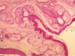
- At the anus, the columnar intestinal epithelium is replaced by the stratified squamous keratinised epithelium of the skin.
- The anal canal joins the bowel to the exterior and is the last 2-3cm of the alimentary tract.
- This is a short passage derived from the proctodeum (formed by invagination of the surface ectoderm).
- Sebaceous and large apocrine sweat glands both occur in this region in association with the anal sphincters.
- Before joining the anal canal, the rectum becomes dilated to form the rectal ampulla.
- At the rectoanal junction, the lumen is constricted by longitudinal folds in the mucosa.
- These are normally pressed together to occlude the lumen.
- As the muscosa changes from columnar to cutaneous, three zones are created:
- Columnar
- Has many longitudinal folds.
- Divided from the rectum by the anorectal line.
- This is a line where the mucosa is stratified squamous epithelium containing lots of lymphoid tissue.
- Intermediate
- Divided from the cutaneous zone by the anocutaneous line.
- Cutaneous
- Skin.
- Stratified squamous keratinised epithelium.
- Surrounds the anus.
- Excretory ducts of the anal sacs open into this region.
- Columnar
Species Differences
Carnivore
- The dog and cat posses two anal sacs. In the dog, these are the size of a hazlenut.
- Located ventrolaterally between the internal and external anal sphincters.
- Large, coiled apocrine tubules that have many tubuloalveolar glands that produce a fatty secretion in their walls.
- The fundus of the sac secretes a potent smelling fluid that drains through a single duct to an opening near the anocutaneous juncntion.
- The anal sacs get compressed during defecation, which causes the fluid to be expressed. The scent of the fluid is thought to act as a territorial marker.
- Anal sacs are clinically important as they are commonly diseased in dogs - frequently, they become enlarged due to accumulated secretion.
- The dog has perianal glands that lie in the skin surrounding the anus.
- They are modified sebaceous glands.
Pig
- Also have anal sacs.

