Difference between revisions of "Coccidiosis - Cattle"
| Line 1: | Line 1: | ||
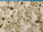
| − | [[Image:Coccidia ruminant.jpg|thumb|right|150px| | + | [[Image:Coccidia ruminant.jpg|thumb|right|150px|<i>Eimeria</i> sp. of ruminants - Joaquim Castellà Veterinary Parasitology Universitat Autònoma de Barcelona]] [[Image:Coccidia oocyst ruminant.jpg|thumb|right|150px|Coccidia oocyst from ruminant faeces - Joaquim Castellà Veterinary Parasitology Universitat Autònoma de Barcelona]] |
| − | [[Image:Coccidia oocyst ruminant.jpg|thumb|right|150px|Coccidia oocyst from ruminant faeces - Joaquim Castellà Veterinary Parasitology Universitat Autònoma de Barcelona]] | ||
| − | |||
| − | + | == Introduction == | |
| − | + | Coccidiosis is primarily a disease of groups of cattle less than one year old. It often occurs if these groups are housed or kept in unhygienic conditions, as like coccidiosis in other species, coccidiosis in cattle is a disease of over-crowding and poor hygiene. Calves with concurrent infections or ones with poor body condition are also susceptable. Bought-in calves that are then mixed with current stock is one of the primary causes of the disease and clinical signs are usually appa<span id="fck_dom_range_temp_1299253210635_717" />rent around a month after this event has occured. | |
| − | + | Infection is usually sporadic, but once immunity has developed it is likely not to reoccur. During the neonatal period, passive immunity is sufficient, only after this wanes are clincal signs of the disease apparent. | |
| − | |||
| − | |||
| − | |||
| − | |||
| − | + | There are many species of coccidia that affect cattle, but Eimeria zuernii, E. bovis and E. alabamensis are by far the most common. E. zuernii is the most pathogenic of these species. | |
| − | |||
| − | |||
| − | |||
| − | |||
| − | |||
| − | |||
| − | |||
| − | + | == Clinical Signs == | |
| − | + | All species of coccidia produce diarrhoea or dysentry with notible tenesmus. | |
| − | |||
| − | |||
| − | |||
| − | |||
| − | |||
| − | + | == Diagnosis == | |
| − | |||
| − | |||
| − | + | History and clinical signs plus husbandry and signalment of animals is indicative of the disease. | |
| − | |||
| − | |||
| − | |||
| − | |||
| − | + | Faecal samples should be taken for faecal floatation and microscopic examination for oocysts. High numbers of oocysts are indicative of clinical disease as small numbers of oocysts are present in all cattle. If the faecal sample is taken after the main oocyst production phase has passed then number will be low even in severely affected animals. | |
| − | |||
| − | + | Post mortem examination will reveal diffuse inflammation and thickening of caecal mucosa (and sometimes ileal and colonic mucosa). Scrapings of the mucosa should be taken and viewed microscopically. In an infected animal masses of gamonts and oocysts will be present in these scrapings. | |
| − | |||
| − | |||
| − | |||
| + | == Treatment and Control == | ||
| − | [[Category:Coccidia]][[Category: | + | As with all causes of diarrhoea, supportive therapy such as rehydration and electrolyte solutions are necessary. |
| − | [[Category:To_Do_- | + | |
| + | Anti-coccidial drugs such as diclazuril or sulphonamides should be started once the diagnosis is confirmed and the affected animals should be moved to a clean environment. All in contact cattle should be treated with the same drug, should costs allow. | ||
| + | |||
| + | Control measures focus on improving husbandry to decrease over-crowding, improve hygiene conditions and prevent mixing of newly purchased stock. | ||
| + | |||
| + | Preventative in- feed medication can be supplied such as decoquinate. | ||
| + | |||
| + | |||
| + | |||
| + | == References == | ||
| + | |||
| + | Andrews, A.H, Blowey, R.W, Boyd, H and Eddy, R.G. (2004) Bovine Medicine (Second edition), Blackwell Publishing <br>Fox, M and Jacobs, D. (2007) Parasitology Study Guide Part 1: Ectoparasites Royal Veterinary College <br>Radostits, O.M, Arundel, J.H, and Gay, C.C. (2000) Veterinary Medicine: a textbook of the diseases of cattle, sheep, pigs, goats and horses Elsevier Health Sciences <br> | ||
| + | |||
| + | |||
| + | |||
| + | Test yourself with the Coccidia Flashcards | ||
| + | |||
| + | [[Coccidia Flashcards|Coccidia Flashcards]] | ||
| + | |||
| + | [[Category:Coccidia]] [[Category:Intestinal_Diseases_-_Cattle]] [[Category:To_Do_-_Review]] | ||
Revision as of 15:58, 4 March 2011
Introduction
Coccidiosis is primarily a disease of groups of cattle less than one year old. It often occurs if these groups are housed or kept in unhygienic conditions, as like coccidiosis in other species, coccidiosis in cattle is a disease of over-crowding and poor hygiene. Calves with concurrent infections or ones with poor body condition are also susceptable. Bought-in calves that are then mixed with current stock is one of the primary causes of the disease and clinical signs are usually apparent around a month after this event has occured.
Infection is usually sporadic, but once immunity has developed it is likely not to reoccur. During the neonatal period, passive immunity is sufficient, only after this wanes are clincal signs of the disease apparent.
There are many species of coccidia that affect cattle, but Eimeria zuernii, E. bovis and E. alabamensis are by far the most common. E. zuernii is the most pathogenic of these species.
Clinical Signs
All species of coccidia produce diarrhoea or dysentry with notible tenesmus.
Diagnosis
History and clinical signs plus husbandry and signalment of animals is indicative of the disease.
Faecal samples should be taken for faecal floatation and microscopic examination for oocysts. High numbers of oocysts are indicative of clinical disease as small numbers of oocysts are present in all cattle. If the faecal sample is taken after the main oocyst production phase has passed then number will be low even in severely affected animals.
Post mortem examination will reveal diffuse inflammation and thickening of caecal mucosa (and sometimes ileal and colonic mucosa). Scrapings of the mucosa should be taken and viewed microscopically. In an infected animal masses of gamonts and oocysts will be present in these scrapings.
Treatment and Control
As with all causes of diarrhoea, supportive therapy such as rehydration and electrolyte solutions are necessary.
Anti-coccidial drugs such as diclazuril or sulphonamides should be started once the diagnosis is confirmed and the affected animals should be moved to a clean environment. All in contact cattle should be treated with the same drug, should costs allow.
Control measures focus on improving husbandry to decrease over-crowding, improve hygiene conditions and prevent mixing of newly purchased stock.
Preventative in- feed medication can be supplied such as decoquinate.
References
Andrews, A.H, Blowey, R.W, Boyd, H and Eddy, R.G. (2004) Bovine Medicine (Second edition), Blackwell Publishing
Fox, M and Jacobs, D. (2007) Parasitology Study Guide Part 1: Ectoparasites Royal Veterinary College
Radostits, O.M, Arundel, J.H, and Gay, C.C. (2000) Veterinary Medicine: a textbook of the diseases of cattle, sheep, pigs, goats and horses Elsevier Health Sciences
Test yourself with the Coccidia Flashcards

