Difference between revisions of "Cyclophyllidea"
m |
|||
| Line 19: | Line 19: | ||
== Introduction == | == Introduction == | ||
| − | *Cyclophyllidean tapeworms have four circular suckers around the scolex. Some species also have hooks on the suckers, and these species are said to be ‘armed’. They also consist of a short unsegmented neck and a chain of segments. | + | *Cyclophyllidean tapeworms have four circular suckers around the scolex (head). Some species also have hooks on the suckers, and these species are said to be ‘armed’. They also consist of a short unsegmented neck and a chain of segments. |
*Most cyclophyllidean species live in the small intestine. New segments bud off from behind the scolex. These do not have an alimentary tract, but absorb nutrients across the body surface. This is covered by a tegument which is like that as for trematodes, but has a microthrix (minute finger-like projections) to increase surface area. Below the tegument are muscle cells and the parenchyma – a syncitium of cells, which fills the space between the organs. The nervous system consists of ganglia in the scolex, from which nerves enter the strobila. The excretory system is composed of flame cells leading to efferent canals, which run through the strobila to discharge at the terminal segment. | *Most cyclophyllidean species live in the small intestine. New segments bud off from behind the scolex. These do not have an alimentary tract, but absorb nutrients across the body surface. This is covered by a tegument which is like that as for trematodes, but has a microthrix (minute finger-like projections) to increase surface area. Below the tegument are muscle cells and the parenchyma – a syncitium of cells, which fills the space between the organs. The nervous system consists of ganglia in the scolex, from which nerves enter the strobila. The excretory system is composed of flame cells leading to efferent canals, which run through the strobila to discharge at the terminal segment. | ||
| Line 26: | Line 26: | ||
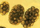
*Outside the body, the eggs are freed by disintegration of the segment, or are shed through the genital pore. Each egg is immediately infective and contains a tapeworm larva with six hooks (the oncosphere), surrounded by a thick, dark ‘shell’ made of numerous blocks (giving a striated appearance when viewed under the microscope). A true shell, which is a delicate membrane, is often lost while still in the uterus. | *Outside the body, the eggs are freed by disintegration of the segment, or are shed through the genital pore. Each egg is immediately infective and contains a tapeworm larva with six hooks (the oncosphere), surrounded by a thick, dark ‘shell’ made of numerous blocks (giving a striated appearance when viewed under the microscope). A true shell, which is a delicate membrane, is often lost while still in the uterus. | ||
| − | |||
== Life-Cycle == | == Life-Cycle == | ||
Revision as of 17:49, 11 January 2010
|
|
Introduction
- Cyclophyllidean tapeworms have four circular suckers around the scolex (head). Some species also have hooks on the suckers, and these species are said to be ‘armed’. They also consist of a short unsegmented neck and a chain of segments.
- Most cyclophyllidean species live in the small intestine. New segments bud off from behind the scolex. These do not have an alimentary tract, but absorb nutrients across the body surface. This is covered by a tegument which is like that as for trematodes, but has a microthrix (minute finger-like projections) to increase surface area. Below the tegument are muscle cells and the parenchyma – a syncitium of cells, which fills the space between the organs. The nervous system consists of ganglia in the scolex, from which nerves enter the strobila. The excretory system is composed of flame cells leading to efferent canals, which run through the strobila to discharge at the terminal segment.
- Each segment develops male and female organs, but they usually cross-fertilise. The genital pores are lateral. The proglottids become sexually mature as they pass down the strobila. As the segment matures, its internal structure largely disappears and the gravid proglottid eventually only contains remnants of the branched uterus packed with eggs. The ‘gravid’ (that is to say, pregnant) segments at the end of the chain may contain greater than 100,000 eggs. In general, one or two segments drop off daily to exit the animal by their own mobility or to be swept out with the faeces. The gravid segments are shed intact from the strobila.
- Outside the body, the eggs are freed by disintegration of the segment, or are shed through the genital pore. Each egg is immediately infective and contains a tapeworm larva with six hooks (the oncosphere), surrounded by a thick, dark ‘shell’ made of numerous blocks (giving a striated appearance when viewed under the microscope). A true shell, which is a delicate membrane, is often lost while still in the uterus.
Life-Cycle
Indirect with one or more intermediate hosts. When the egg is ingested by the intermediate host, the gastric and intestinal secretions digest the thick shell and activate the 6-hooked oncosphere. Using its hooks, it tears through the mucosa of the host to reach the blood or lymph system, or in the case of invertebrates, the body cavity. Once it has reached its predilection site, the oncosphere loses its hooks and develops, depending on the species, into one of the following larval stages, known as a metacestode. There are six types of metacestode (in increasing order of complexity:
1) Cysticercus: a fluid-filled bladder with one inverted scolex
2) Cysticercoid: pinhead size; only found in invertebrates; like the cysticercus, but the bladder is reduced to a potential space and the scolex is not inverted
3) Strobilocercus: restricted to the cat tapeworm Taenia taeniaeformis; like a cysticercus, but the single scolex is attached to the bladder by a chain of segments
4) Coenurus: like a cysticercus, but has multiple inverted scolices
5) Hydatid cyst: the metacestode of Echinococcus granulosus; this fluid-filled bladder can grow to the size of a football; it is lined with germinal epithelium that buds off brood capsules internally; inverted scolices form inside these; hydatid sand is the name given to the brood capsules and scolices in the hydatid fluid; the host attempts to wall off the hydatid cyst with fibrous tissue; between this and the germinal membrane is an amorphous layer
6) Alveolar cyst: the metacestode of Echinococcus multilocularis; this is like the hydatid cyst, but daughter cysts bud off the external, as well as the internal, surface of the germinal layer, with the result that the metacestode expands by infiltrating through the tissue, rather like a tumour.
When the metacestode is ingested by the final host, the scolex attaches to the mucosa, the remainder of the structure is digested, and a chain of proglottids (segments) begins to grow from the base of the scolex.
Infection of the final-host involves at least three epidemiological relationships:
1) predator-prey, e.g. cat eating infected mouse
2) accidental, e.g. horse eating infected pasture mites
3) irritation, e.g. infected flea on animal = exaggerated grooming of animal = swallowed

