Difference between revisions of "Cyclophyllidea Life-Cycle"
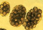
(Created page with 'right|150px|thumb|''Dipylidium caninum'' - Courtesy of the Laboratory of Parasitology, University of Pennsylvania School of Veterinary Medicine…') |
|||
| Line 22: | Line 22: | ||
2) '''accidental''', e.g. horse eating infected pasture mites | 2) '''accidental''', e.g. horse eating infected pasture mites | ||
| − | 3) '''irritation''', e.g. infected flea on animal = exaggerated grooming of animal = swallowed[[Category:Cyclophyllidea]] | + | 3) '''irritation''', e.g. infected flea on animal = exaggerated grooming of animal = swallowed |
| + | |||
| + | [[Category:Cyclophyllidea]][[Category:To_Do_-_Parasites]] | ||
Revision as of 21:51, 25 June 2010
Indirect with one or more intermediate hosts. When the egg is ingested by the intermediate host, the gastric and intestinal secretions digest the thick shell and activate the 6-hooked oncosphere. Using its hooks, it tears through the mucosa of the host to reach the blood or lymph system, or in the case of invertebrates, the body cavity. Once it has reached its predilection site, the oncosphere loses its hooks and develops, depending on the species, into one of the following larval stages, known as a metacestode. There are six types of metacestode (in increasing order of complexity:
1) Cysticercus: a fluid-filled bladder with one inverted scolex
2) Cysticercoid: pinhead size; only found in invertebrates; like the cysticercus, but the bladder is reduced to a potential space and the scolex is not inverted
3) Strobilocercus: restricted to the cat tapeworm Taenia taeniaeformis; like a cysticercus, but the single scolex is attached to the bladder by a chain of segments
4) Coenurus: like a cysticercus, but has multiple inverted scolices
5) Hydatid cyst: the metacestode of Echinococcus granulosus; this fluid-filled bladder can grow to the size of a football; it is lined with germinal epithelium that buds off brood capsules internally; inverted scolices form inside these; hydatid sand is the name given to the brood capsules and scolices in the hydatid fluid; the host attempts to wall off the hydatid cyst with fibrous tissue; between this and the germinal membrane is an amorphous layer
6) Alveolar cyst: the metacestode of Echinococcus multilocularis; this is like the hydatid cyst, but daughter cysts bud off the external, as well as the internal, surface of the germinal layer, with the result that the metacestode expands by infiltrating through the tissue, rather like a tumour.
When the metacestode is ingested by the final host, the scolex attaches to the mucosa, the remainder of the structure is digested, and a chain of proglottids (segments) begins to grow from the base of the scolex.
Infection of the final-host involves at least three epidemiological relationships:
1) predator-prey, e.g. cat eating infected mouse
2) accidental, e.g. horse eating infected pasture mites
3) irritation, e.g. infected flea on animal = exaggerated grooming of animal = swallowed
