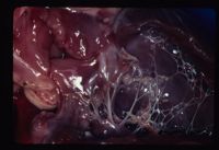
Degenerative Mitral Valve Disease
| This article is still under construction. |
Also known as: MVD, Mitral insufficiency, Mitral endocardiosis, Myxomatous Mitral Valve Disease (MMVD)
- Common in dogs and cats
- Rare in other species
Signalment
Typically in middle aged to older small breed dogs. Genetically predisposed breeds include Cavalier King Charles Spaniel, Bull Terriers, German Shepherds, and Great Danes.
Introduction
A congenital malformation or degeneration of the mitral valve leaflets and its supporting structures(chordae tendineae, papillary muscles, valvular leaflets, annulus) results in valvular regurgitation (insufficiency)
Chronic mitral regurgitation leads to volume overload of the left heart, which results in dilatation (eccentric hypertrophy) of the left ventricle and atrium. When mitral regurgitation is severe, cardiac output decreases, which results in signs of left sided cardiac failure LCHF and pulmonary venous congestion. Dilatation of the left-sided chambers predisposes affected animals to arrhythmias. In some cases, malformation of the mitral valve complex causes a degree of valvular stenosis as well as insufficiency.
Diagnosis
History
Clinical Signs
Diagnostic imaging
Laboratory Tests
--- Mild defects may be asymptomatic
Signs of left sided congestive (dilated) heart failure:
-Cough
-Tachypnea/Dyspnea
-Exercise Intolerance
-Syncope
-Pale Mucous Membranes
-Tachycardia
-Arrhythmias
Physical Exam
-Left apical systolic murmur
-Left diastolic murmur
-Poor pulses
Radiographic Findings
-Left atrial enlargement
-Left ventricular enlargement
-Enlargement of pulmonary veins
-Pulmonary edema
Echocardiographic Findings
-Left atrial enlargement
-Left ventricular enlargement
-Malformed valve leaflets
Doppler can detect mitral stenosis or regurgitation and estimate pressures in the left atrium and pulmonary veins
Electrocardiographic (ECG)
-May be normal
-Signs of left atrial (wide P waves)
-Signs of left ventricular (tall R waves)
-Signs of arrhythmias
Treatment
Control left atrial pressure and manage left sided congestive heart failure
Goal of treatment for congestive heart failure:
-Diuretics (decrease venous congestion)
-ACE-inhibitors; Vasodilators (inhibit water retention and dilate the vessels)
-Anti-coagulants (cats) to prevent thrombus formation
Prognosis
Mild Defects
-Excellent
Severe Defects
-Guarded
From Pathology
Often associated with mitral regurgitation and left atrial volume overload. Usually progresses to left sided heart failure.
Incidence:
- Most common congenital defect in cats.
- Also reported in pure breed dogs E.g GSD, Great Danes.
Clinical Signs:
- Often murmur is the only clinical sign; pansystolic with increased intensity over the mitral valve area.
- May also see exercise intolerance, dyspnoea and coughing.
Diagnosis:
- Left atrial enlargement on radiology and ECG.
- Doppler echocardiography can dtect abnormal flow.
Treatment:
- Prognosis poor, medically manage left heart failure with ACE-inhibitors etc.
