Difference between revisions of "Electrocardiography"
| Line 1: | Line 1: | ||
| − | |||
{{toplink | {{toplink | ||
|linkpage =Anaesthesia | |linkpage =Anaesthesia | ||
| Line 8: | Line 7: | ||
|pagetype=Clinical | |pagetype=Clinical | ||
}} | }} | ||
| − | |||
==Introduction== | ==Introduction== | ||
Electrocardiography is one of the most commonly found piece of monitoring equipment in modern veterinary practices. It detects the electrical activity of the heart through 3 electrodes. These electrodes are most commonly placed on the 2 forelimbs and the left hindlimb. The electrodes are attached to the patient via ECG pads (most commonly), crocodile clips (more common in horses) and transcutaneous needles (rare). Frequently, additional electrode gel or alcohol is required to improve contact between the patient and electrodes. | Electrocardiography is one of the most commonly found piece of monitoring equipment in modern veterinary practices. It detects the electrical activity of the heart through 3 electrodes. These electrodes are most commonly placed on the 2 forelimbs and the left hindlimb. The electrodes are attached to the patient via ECG pads (most commonly), crocodile clips (more common in horses) and transcutaneous needles (rare). Frequently, additional electrode gel or alcohol is required to improve contact between the patient and electrodes. | ||
| − | |||
==Reading an ECG Trace== | ==Reading an ECG Trace== | ||
| Line 18: | Line 15: | ||
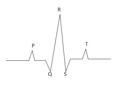
[[Image:ECG.jpg|left|]] | [[Image:ECG.jpg|left|]] | ||
| − | |||
| − | |||
| − | |||
<center> | <center> | ||
| Line 39: | Line 33: | ||
|} | |} | ||
</center> | </center> | ||
| − | + | When interpreting ECG traces it is important to remember the following rules so that arrhythmias can be detected early and treated if necessary; | |
| − | |||
| − | |||
| − | |||
| − | |||
| − | |||
| − | |||
| − | |||
| − | |||
| − | When interpreting ECG traces it is important to remember the following rules so that arrhythmias can be detected early and treated if necessary | ||
| − | |||
*Is there a P for every QRS? | *Is there a P for every QRS? | ||
*Is there a QRS for every P? | *Is there a QRS for every P? | ||
*Are they all reasonably related? | *Are they all reasonably related? | ||
*Are they all the same? | *Are they all the same? | ||
Revision as of 10:37, 8 September 2010
|
|
Introduction
Electrocardiography is one of the most commonly found piece of monitoring equipment in modern veterinary practices. It detects the electrical activity of the heart through 3 electrodes. These electrodes are most commonly placed on the 2 forelimbs and the left hindlimb. The electrodes are attached to the patient via ECG pads (most commonly), crocodile clips (more common in horses) and transcutaneous needles (rare). Frequently, additional electrode gel or alcohol is required to improve contact between the patient and electrodes.
Reading an ECG Trace
An ECG supplies information about the electrical activity of the heart only. It indicates the heart rate and rhythm and can be used to detect any arrhythmias. It does not supply information about cardiac function
| Stage | Represents |
|---|---|
| P | Atrial Depolarisation |
| QRS | Ventricular Depolarisation |
| T | Ventricular Repolarisation |
When interpreting ECG traces it is important to remember the following rules so that arrhythmias can be detected early and treated if necessary;
- Is there a P for every QRS?
- Is there a QRS for every P?
- Are they all reasonably related?
- Are they all the same?
