Difference between revisions of "Gastric Dilatation and Volvulus"
TestStudent (talk | contribs) |
|||
| (61 intermediate revisions by 7 users not shown) | |||
| Line 1: | Line 1: | ||
| − | {{ | + | {{unfinished}} |
| − | |||
| − | == | + | ==Signalment== |
| − | Gastric | + | *Large deep chested breeds including: |
| + | <gallery> | ||
| + | Image:Akita.jpg|Akitas | ||
| + | Image:Bloodhound.jpg|Bloodhounds | ||
| + | Image:Smooth Collie.jpg|Collies | ||
| + | Image:Greatdane.jpg|Great Danes | ||
| + | Image:Irish Setter.jpg|Irish Setters | ||
| + | Image:Irish Wolfhound.jpg|Wolfhounds | ||
| + | Image:Newfoundland.jpg|Newfoundlands | ||
| + | Image:Rottweiler.jpg|Rottweilers | ||
| + | Image:Stbernard.jpg|Saint Bernards | ||
| + | Image:Standard poodle.jpg|Standard Poodles | ||
| + | Image:Weimaraner.jpg|Weirmaraners | ||
| + | </gallery> | ||
| + | |||
| + | ==Description== | ||
| + | Gastric dilatation (GD) and Gastric dilatation and volvulus (GDV) are caused by the stomach distending with air. In GDV the stomach twists around its axis mainly in a clockwise direction with both conditions leading to compression of the caudal vena cava. GDV can lead to [[Shock - Pathology#Hypovolaemic shock|hypovolaemic shock]], splenic torsion, gastric wall ischaemia, abdominal viscera congestion, [[Shock - Pathology#Endotoxic shock|endotoxic shock]] and [[Disseminated Intravascular Coagulation|disseminated intravascular coagulation (DIC)]]. The exact pathogenesis is unclear but risk factors for GDV include age, fast eating, once- daily feeding, aerophagia, raised feeding bowl and a close relative with GDV. | ||
| − | + | ==Diagnosis== | |
| + | ===History and Clinical signs=== | ||
| + | *Abdominal distension | ||
| + | *Non-productive retching | ||
| + | *Weakness | ||
| + | *Collapse | ||
| + | *Salivation | ||
| + | *Abdominal tympany | ||
| + | *Tachycardia | ||
| + | *Pallor | ||
| + | *Hypothermia | ||
| + | *[[:Category:Arrhythmia|Cardiac arrythmias]] (present in 40-50% of patients) ([[Ventricular Premature Complexes|ventricular premature beats]], [[Ventricular Tachycardia|ventricular tachycardia]]) | ||
| − | == | + | ===Haematology=== |
| − | + | *Increased haematocrit | |
| + | *DIC (thrombocytopaenia, increased firbin degradation products, prolonged patial thromboplastin time and reduced antithrombin III.) | ||
| − | == | + | ===Biochemistry=== |
| − | + | Most commonly find hypokalaemia and metabolic acidosis. The acidosis is caused hypoperfusion and anaerobic metabolism leading to lactic acid accumulation. Respiratory acidosis and alkalosis may also be present due to hypo- and hyperventilation. | |
| − | + | ===Diagnostic imaging=== | |
| + | Best performed after [[Principles of Fluid Therapy|fluid therapy]] and gastric decompression. It allows distinction between GD and GDV: | ||
| + | *Gastric dilatation: gas distension, on right lateral shows air in the fundus. | ||
| + | *Gastric dilatation and volvulus: pylorus moves dorsally and left with a compartmentalized stomach. (GDV x-ray from WikiCommons[[http://commons.wikimedia.org/wiki/File:GDV_x-ray.JPG]]) | ||
| + | A right lateral view will show a large fundus ventrally, with a smaller gas filled pylorus located dorsally to that. These are seperated by a soft tissue strip. The contrast of the abdomen may be lost indicating peritonitis or [[Haemoabdomen|haemoabdomen]]. Gastric rupture would show as pneumoperitoneum and increased contrast. | ||
| − | + | ==Treatment== | |
| + | The most important first line treatments are [[Principles of Fluid Therapy|fluid therapy]] and gastric decompression. | ||
| − | == | + | ===Fluid therapy=== |
| − | + | Should be individualised to the patient due to the varying nature of the [[Essential Ion and Compound Balance and Homeostasis - Anatomy & Physiology#Acid / Base|acid-base]] disturbances. Large bore (16 or 18 gauge) catheters should be placed into cephalic or jugular veins (ideally two into both cephalic veins). Shock doses of Compound Sodium Lactate (Lactated Ringer's Solution) (60-90ml/kg/h). Hypertonic saline can also be used. Monitoring of the situation should be done by regular blood pressure measurements, heart rates, PCV and total solids and urine output. Potassium can be supplemented to bags in the form of KCl after the initial shock doses. | |
| − | == | + | ===Gastric decompression=== |
| − | + | Performed by introduction of a lubricated premeasured (from nostril to last rib) stomach tube or by trocharizing the most tympanic area caudal to the ribs with a 14 to 16 gauge catheter. Sedation may be required to allow the passage of the stomach tube. Suitable drugs for this include [[Opioids#Butorphanol|butorphanol]], [[Opioids#Fentanyl|fentanyl]] or oxymorphone and [[Benzodiazepines#Diazepam|diazepam]]. | |
| − | ===[[: | + | ===Other treatment=== |
| − | + | *For shock: [[Steroids|Prednisolone sodium succinate]] or [[Steroids|dexamethasone sodium phosphate]]. | |
| − | + | *For bacterial translocation and endotoxaemia: Broad spectrum antibiotics (e.g. [[Cephalosporins|cephalosporin]] and a [[Fluoroquinolones|fluoroquinolone]]) should also be given at surgical induction through to the postoperative period. | |
| + | *For cardiac arrythmias: indicated if weakness, syncope, tachycardia runs with R on T complexes, ventricular tachycardia at rates >150bpm. Treated by correcting [[Essential Ion and Compound Balance and Homeostasis - Anatomy & Physiology#Acid / Base|acid-base]], [[Essential Ion and Compound Balance and Homeostasis - Anatomy & Physiology|electrolyte]] and [[Haemostasis - Pathology|haemostatic]] disturbances. The treatment is [[Local Anaesthetics#Lidocaine|lidocaine]] by bolus or continuous rate infusion or procainamide if they persist. | ||
| + | *For analgesia: Pure [[Opioids|opioid]] of [[Opioids#Morphine|morphine]], [[Opioids#Methadone|methadone]] or [[Opioids#Fentanyl|fentanyl]]. | ||
| + | *General: Oxygen supplementation if possible | ||
| − | === | + | ===Anaesthesia=== |
| − | + | Anaesthesia must be carried out with care even after the patient has been stabilised. There are limited protocols but included [[Opioids#Fentanyl|fentanyl]] and [[Benzodiazepines#Diazepam|diazepam]] bolus or titrated [[Propofol|propofol]]. Maintenance can be achieved with the use of [[Isoflurane|isoflurane]] and [[Sevoflurane|sevoflurane]] in oxygen however [[Nitrous Oxide|nitrous oxide]] should be avoided due to third spacing. Regular routine monitoring of urine production, blood pressure, [[Blood Pressure#Central Venous Pressure|central venous pressure]], PCV, total solids, blood gas and serum electrolytes. High rates of fluids should be used to maintain tissue perfusion and arterial [[Blood Pressure|blood pressure]]. | |
| − | === | + | ===Surgery=== |
| − | + | Surgical aims include: | |
| + | *Gastric decompression and repositioning | ||
| + | *Assessing the organ viability | ||
| + | *Removing necrotic tissue | ||
| + | *Gastropexy (can perform incisional, tube, belt-loop and circumcostal techniques) to prevent recurrence | ||
| − | + | If gastric necrosis (happens in 10-37% of patients) is present (discoloured dark purple or grey/green, don't bleed when incised or feel paper thin) then a parital gastrectomy is required. Damage to the spleen via avulsion or torsion may need partial or complete splenectomy. | |
| − | |||
| − | + | ===Post-operative complications=== | |
| + | These are wide and varied and include: | ||
| + | *Hypoperfusion | ||
| + | *Hypotension | ||
| + | *Cardiac arrythmias | ||
| + | *[[Lungs Inflammatory - Pathology#Aspiration pneumonia|Aspiration pneumonia]] | ||
| + | *[[Gastric Motility Disorders|Abnormal gastric motility]] | ||
| + | *Gastric necrosis | ||
| + | *[[Disseminated Intravascular Coagulation|DIC]] | ||
| + | *Systemic Inflammatory Response Syndrome (SIRS) | ||
| − | == | + | ==Prognosis== |
| − | + | Simple GDV mortality rates are around 15%. Patients suffering from gastric necrosis, gastric resection or splenectomy have a higher mortality rate at over 30%. Gastric necorsis can be predicted by measuring plasma lactate. Values >6mmol/l indicates necrosis (Specificity 88%, Sensitivity 66%) | |
| + | |||
| + | ==References== | ||
| + | |||
| + | Hall, E.J, Simpson, J.W. and Williams, D.A. (2005) '''BSAVA Manual of Canine and Feline Gastroenterology (2nd Edition)''' ''BSAVA'' | ||
| + | |||
| + | King, L. and Hammond, R. (1999) '''BSAVA Manual of Canine and Feline Emergency and Critical Care''' ''BSAVA'' | ||
| − | + | Tivers, M. and Brockman, D. (2009) '''[[http://inpractice.bvapublications.com/cgi/reprint/31/2/66?maxtoshow=&HITS=10&hits=10&RESULTFORMAT=&author1=brockman&searchid=1&FIRSTINDEX=0&sortspec=relevance&resourcetype=HWCIT|Gastric dilation–volvulus syndrome in dogs 1. Pathophysiology, diagnosis and stabilisation]]''' 31(2):66 ''In Practice'' | |
| − | + | Tivers, M. and Brockman, D. (2009) '''[[http://inpractice.bvapublications.com/cgi/reprint/31/3/114?maxtoshow=&HITS=10&hits=10&RESULTFORMAT=&author1=brockman&andorexactfulltext=and&searchid=1&FIRSTINDEX=0&sortspec=relevance&resourcetype=HWCIT|Gastric dilation–volvulus syndromein dogs 2. Surgical and postoperative management]]''' 31(3):114 ''In Practice'' | |
| − | + | ==From pathology section== | |
| − | + | * Is a consequence of '''gastric dilation'''. | |
| + | ** Gastric dilation occurs in dogs, cats, horses, rabbits and primates. | ||
| + | ** Cause unclear but may be associated with overeating. | ||
| + | ** Gastric dilation is most studied in dog, since it can lead to displacement of the [[Forestomach - Anatomy & Physiology|stomach]] within the abdomen. | ||
| − | + | ===Clinical=== | |
| − | + | * Mainly affects large, deep-chested dogs - Great Dane, St. Bernard's and occasionally German Shepherd dogs. | |
| − | + | ** A similar condition also occurs in the pig. | |
| + | * Animal collapses suddenly and must be operated on rapidly. | ||
| − | == | + | ===Pathogenesis=== |
| − | + | ||
| + | * Usually occurs around 30 minutes after a meal, or following aerophagia. | ||
| + | ** [[Forestomach - Anatomy & Physiology|Stomach]] is distended (gastric dilation). | ||
| + | ** Animal excercises, and the [[Forestomach - Anatomy & Physiology|stomach]] twists 180 degrees clockwise on its mesentery. | ||
| + | * Torsion impairs the blood supply- the arterial supply is maintained BUT venous drainage is blocked. | ||
| + | ** [[Forestomach - Anatomy & Physiology|Stomach]] wall becomes severely congested and infarction of gastric mucosa may occur. | ||
| + | * [[Forestomach - Anatomy & Physiology|Stomach]] blows up with gas and fluid. | ||
| + | ** Block venous return to heart. | ||
| + | ** Compresses diaphragm and interferes with respiration. | ||
| − | + | * '''The actual cause of the problem and the reason for accumulation of gas is unclear.''' | |
| − | + | ** It is better to feed big dogs small amounts more frequently. | |
| − | |||
| − | |||
| − | |||
| − | == | + | ===Pathology=== |
| − | |||
| − | |||
| − | |||
| − | |||
| − | |||
| − | |||
| − | |||
| + | ====Gross==== | ||
| − | + | * Following the pathogenesis above the [[Forestomach - Anatomy & Physiology|stomach]] contents appear dark red/black and bloody, and the organ may rupture. | |
| + | * The [[Spleen - Anatomy & Physiology|spleen]] is also affected by venous occlusion. | ||
| + | ** Becomes very congested and moves from left to right side of abdomen. | ||
| − | + | ====Histological==== | |
| − | [[Category:Stomach_and_Abomasum_-_Pathology]][[Category: | + | * Venous obstruction gives rise to congestion, oedema and necrosis of gastric mucosa.[[Category:Stomach_and_Abomasum_-_Pathology]][[Category:Dog]] |
| − | [[Category: | + | [[Category:To_Do_-_Stuart]] |
Revision as of 16:13, 5 July 2010
| This article is still under construction. |
Signalment
- Large deep chested breeds including:
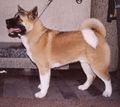
Akitas
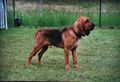
Bloodhounds
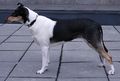
Collies
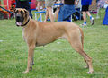
Great Danes
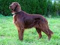
Irish Setters
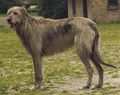
Wolfhounds
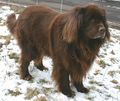
Newfoundlands
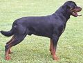
Rottweilers
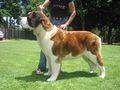
Saint Bernards
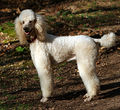
Standard Poodles
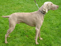
Weirmaraners


Description
Gastric dilatation (GD) and Gastric dilatation and volvulus (GDV) are caused by the stomach distending with air. In GDV the stomach twists around its axis mainly in a clockwise direction with both conditions leading to compression of the caudal vena cava. GDV can lead to hypovolaemic shock, splenic torsion, gastric wall ischaemia, abdominal viscera congestion, endotoxic shock and disseminated intravascular coagulation (DIC). The exact pathogenesis is unclear but risk factors for GDV include age, fast eating, once- daily feeding, aerophagia, raised feeding bowl and a close relative with GDV.
Diagnosis
History and Clinical signs
- Abdominal distension
- Non-productive retching
- Weakness
- Collapse
- Salivation
- Abdominal tympany
- Tachycardia
- Pallor
- Hypothermia
- Cardiac arrythmias (present in 40-50% of patients) (ventricular premature beats, ventricular tachycardia)
Haematology
- Increased haematocrit
- DIC (thrombocytopaenia, increased firbin degradation products, prolonged patial thromboplastin time and reduced antithrombin III.)
Biochemistry
Most commonly find hypokalaemia and metabolic acidosis. The acidosis is caused hypoperfusion and anaerobic metabolism leading to lactic acid accumulation. Respiratory acidosis and alkalosis may also be present due to hypo- and hyperventilation.
Diagnostic imaging
Best performed after fluid therapy and gastric decompression. It allows distinction between GD and GDV:
- Gastric dilatation: gas distension, on right lateral shows air in the fundus.
- Gastric dilatation and volvulus: pylorus moves dorsally and left with a compartmentalized stomach. (GDV x-ray from WikiCommons[[1]])
A right lateral view will show a large fundus ventrally, with a smaller gas filled pylorus located dorsally to that. These are seperated by a soft tissue strip. The contrast of the abdomen may be lost indicating peritonitis or haemoabdomen. Gastric rupture would show as pneumoperitoneum and increased contrast.
Treatment
The most important first line treatments are fluid therapy and gastric decompression.
Fluid therapy
Should be individualised to the patient due to the varying nature of the acid-base disturbances. Large bore (16 or 18 gauge) catheters should be placed into cephalic or jugular veins (ideally two into both cephalic veins). Shock doses of Compound Sodium Lactate (Lactated Ringer's Solution) (60-90ml/kg/h). Hypertonic saline can also be used. Monitoring of the situation should be done by regular blood pressure measurements, heart rates, PCV and total solids and urine output. Potassium can be supplemented to bags in the form of KCl after the initial shock doses.
Gastric decompression
Performed by introduction of a lubricated premeasured (from nostril to last rib) stomach tube or by trocharizing the most tympanic area caudal to the ribs with a 14 to 16 gauge catheter. Sedation may be required to allow the passage of the stomach tube. Suitable drugs for this include butorphanol, fentanyl or oxymorphone and diazepam.
Other treatment
- For shock: Prednisolone sodium succinate or dexamethasone sodium phosphate.
- For bacterial translocation and endotoxaemia: Broad spectrum antibiotics (e.g. cephalosporin and a fluoroquinolone) should also be given at surgical induction through to the postoperative period.
- For cardiac arrythmias: indicated if weakness, syncope, tachycardia runs with R on T complexes, ventricular tachycardia at rates >150bpm. Treated by correcting acid-base, electrolyte and haemostatic disturbances. The treatment is lidocaine by bolus or continuous rate infusion or procainamide if they persist.
- For analgesia: Pure opioid of morphine, methadone or fentanyl.
- General: Oxygen supplementation if possible
Anaesthesia
Anaesthesia must be carried out with care even after the patient has been stabilised. There are limited protocols but included fentanyl and diazepam bolus or titrated propofol. Maintenance can be achieved with the use of isoflurane and sevoflurane in oxygen however nitrous oxide should be avoided due to third spacing. Regular routine monitoring of urine production, blood pressure, central venous pressure, PCV, total solids, blood gas and serum electrolytes. High rates of fluids should be used to maintain tissue perfusion and arterial blood pressure.
Surgery
Surgical aims include:
- Gastric decompression and repositioning
- Assessing the organ viability
- Removing necrotic tissue
- Gastropexy (can perform incisional, tube, belt-loop and circumcostal techniques) to prevent recurrence
If gastric necrosis (happens in 10-37% of patients) is present (discoloured dark purple or grey/green, don't bleed when incised or feel paper thin) then a parital gastrectomy is required. Damage to the spleen via avulsion or torsion may need partial or complete splenectomy.
Post-operative complications
These are wide and varied and include:
- Hypoperfusion
- Hypotension
- Cardiac arrythmias
- Aspiration pneumonia
- Abnormal gastric motility
- Gastric necrosis
- DIC
- Systemic Inflammatory Response Syndrome (SIRS)
Prognosis
Simple GDV mortality rates are around 15%. Patients suffering from gastric necrosis, gastric resection or splenectomy have a higher mortality rate at over 30%. Gastric necorsis can be predicted by measuring plasma lactate. Values >6mmol/l indicates necrosis (Specificity 88%, Sensitivity 66%)
References
Hall, E.J, Simpson, J.W. and Williams, D.A. (2005) BSAVA Manual of Canine and Feline Gastroenterology (2nd Edition) BSAVA
King, L. and Hammond, R. (1999) BSAVA Manual of Canine and Feline Emergency and Critical Care BSAVA
Tivers, M. and Brockman, D. (2009) [dilation–volvulus syndrome in dogs 1. Pathophysiology, diagnosis and stabilisation] 31(2):66 In Practice
Tivers, M. and Brockman, D. (2009) [dilation–volvulus syndromein dogs 2. Surgical and postoperative management] 31(3):114 In Practice
From pathology section
- Is a consequence of gastric dilation.
- Gastric dilation occurs in dogs, cats, horses, rabbits and primates.
- Cause unclear but may be associated with overeating.
- Gastric dilation is most studied in dog, since it can lead to displacement of the stomach within the abdomen.
Clinical
- Mainly affects large, deep-chested dogs - Great Dane, St. Bernard's and occasionally German Shepherd dogs.
- A similar condition also occurs in the pig.
- Animal collapses suddenly and must be operated on rapidly.
Pathogenesis
- Usually occurs around 30 minutes after a meal, or following aerophagia.
- Torsion impairs the blood supply- the arterial supply is maintained BUT venous drainage is blocked.
- Stomach wall becomes severely congested and infarction of gastric mucosa may occur.
- Stomach blows up with gas and fluid.
- Block venous return to heart.
- Compresses diaphragm and interferes with respiration.
- The actual cause of the problem and the reason for accumulation of gas is unclear.
- It is better to feed big dogs small amounts more frequently.
Pathology
Gross
- Following the pathogenesis above the stomach contents appear dark red/black and bloody, and the organ may rupture.
- The spleen is also affected by venous occlusion.
- Becomes very congested and moves from left to right side of abdomen.
Histological
- Venous obstruction gives rise to congestion, oedema and necrosis of gastric mucosa.