Difference between revisions of "Gingival Hyperplasia"
m (Text replace - "[[Crown|" to "[[Enamel Organ#Crown|") |
|||
| Line 1: | Line 1: | ||
| − | == | + | == Introduciton == |
| − | + | Gingival hyperplasia often appears as pink, hyperaemic and ulcerated lesions that can be either firm or soft. There can be varying amounts of pigmentation reflecting the normal pigmentation of the oral mucosa. [[Enamel Organ#Crown|Crowns]] of teeth are often partially or completely covered by the hyperplastic gingiva forming a potential space or pocket between the gingiva and the [[Enamel Organ#Crown|crown]] where plaque is able to accumulate. | |
| − | + | <br> | |
| + | |||
| + | Gingival hyperplasia can be described as focal lesions, multiple focal lesions or generalised lesions; or a combination of all of these. | ||
| + | <br> | ||
| + | |||
| + | It is thought to be the result of an imbalance in the plaque/host tissue response. There are many factors that can cause this condition. These include drugs such as cicliosporin, phenytoin and calcium channel blockers. Chronic irritation and dental plaque are also causative. Other causes include odontoplastic resorptive lesions, neoplasia and mechanical irritation. | ||
| + | |||
| + | <br> | ||
| + | |||
| + | == Signalment == | ||
| + | This is a common condition in dogs but less common in cats. The following breeds are predisposed: | ||
| Line 11: | Line 21: | ||
</gallery> | </gallery> | ||
| − | + | <br> | |
| − | |||
| − | |||
| − | |||
| − | |||
| − | |||
| − | |||
| − | |||
| − | |||
| − | |||
| − | |||
| − | |||
| − | |||
| − | |||
| − | |||
| − | |||
| − | |||
| − | |||
| − | |||
| + | == Clinical Signs == | ||
| + | Signs depend on the severity of gingival hyperplasia and the degree to which the teeth are covered. They inclide pain on mastication, drooling +/- blood in saliva, Haemorrhage of the ginigiva, reluctance to eat and dysphagia. The animal may paw its mouth or rub its mouth along the floor. | ||
| − | + | <br> | |
| − | == | + | == Diagnosis == |
| − | + | Clinical signs are indicative of the condition. A detailed oral examination under sedation will lead to a presumptive diagnosis. | |
| − | + | <br> | |
| − | |||
| − | |||
| − | |||
| − | |||
===Diagnostic Imaging=== | ===Diagnostic Imaging=== | ||
Oral radiographs should be taken to rule out concurrent conditions. One such condition is periodontitis which is demonstrated radiographically by alveolar bone loss associated with pocket formation between the tooth crown and gingiva. | Oral radiographs should be taken to rule out concurrent conditions. One such condition is periodontitis which is demonstrated radiographically by alveolar bone loss associated with pocket formation between the tooth crown and gingiva. | ||
| + | <br> | ||
===Biopsy=== | ===Biopsy=== | ||
Biopsy samples should include those areas of [[Gingiva Introduction|gingiva]] that show signs of inflammation with a softer than normal texture. Any gingiva with radiographic signs of bone involvement should also be sampled. | Biopsy samples should include those areas of [[Gingiva Introduction|gingiva]] that show signs of inflammation with a softer than normal texture. Any gingiva with radiographic signs of bone involvement should also be sampled. | ||
| − | + | <br> | |
| + | == Treatment == | ||
The suspected cause of the condition should be corrected first. This may include a multimodal treatment plan aimed at controlling plaque formation including teeth brushing and providing the animal with sticks/toys that clean the teeth crowns. | The suspected cause of the condition should be corrected first. This may include a multimodal treatment plan aimed at controlling plaque formation including teeth brushing and providing the animal with sticks/toys that clean the teeth crowns. | ||
| + | <br> | ||
| + | '''Gingivectomy and gingivoplasty''' should be carried out under general anaesthetic if significant pseudo-pockets are present between the gingiva and teeth crowns. The aim should be to eliminate the pseudopockets and re-establish the normal anatomy of the gingival margin. | ||
| + | <br> | ||
| + | Electrosurgery and Laser surgery can be performed. Care must be taken with electrosurgery to avoid contact between the teeth crowns and the electrodes to prevent irreversible heat damage to the pulp. | ||
| − | + | <br> | |
| − | |||
| − | ==Prognosis== | + | == Prognosis == |
The prognosis following surgical excision and histopathology is good. However, local recurrence is possible but less common if a treatment plan aimed at reducing plaque formation is implemented. A re-examination of the patient should be carried out at least every 6 months to assess for signs of recurrence and the sufficiency of plaque control measures. | The prognosis following surgical excision and histopathology is good. However, local recurrence is possible but less common if a treatment plan aimed at reducing plaque formation is implemented. A re-examination of the patient should be carried out at least every 6 months to assess for signs of recurrence and the sufficiency of plaque control measures. | ||
| + | |||
| + | <br> | ||
==References== | ==References== | ||
| + | Tutt, C., Deeprose, J. and Crossley, D. (2007) BSAVA Manual of Canine and Feline Dentistry (3rd Edition), British Small Animal Veterinary Association. | ||
| + | <br> | ||
| + | Merck & Co (2008) The Merck Veterinary Manual, Merial. | ||
| − | |||
| − | + | [[Category:Oral_Cavity_-_Proliferative_Pathology]][[Category:Teeth_-_Proliferative_Pathology]][[Category:To_Do_-_Review]] | |
| − | [[Category:Oral_Cavity_-_Proliferative_Pathology]][[Category:Teeth_-_Proliferative_Pathology]][[Category:To_Do_- | ||
[[Category:Oral Diseases - Dog]][[Category:Oral Diseases - Cat]] | [[Category:Oral Diseases - Dog]][[Category:Oral Diseases - Cat]] | ||
Revision as of 11:28, 30 March 2011
Introduciton
Gingival hyperplasia often appears as pink, hyperaemic and ulcerated lesions that can be either firm or soft. There can be varying amounts of pigmentation reflecting the normal pigmentation of the oral mucosa. Crowns of teeth are often partially or completely covered by the hyperplastic gingiva forming a potential space or pocket between the gingiva and the crown where plaque is able to accumulate.
Gingival hyperplasia can be described as focal lesions, multiple focal lesions or generalised lesions; or a combination of all of these.
It is thought to be the result of an imbalance in the plaque/host tissue response. There are many factors that can cause this condition. These include drugs such as cicliosporin, phenytoin and calcium channel blockers. Chronic irritation and dental plaque are also causative. Other causes include odontoplastic resorptive lesions, neoplasia and mechanical irritation.
Signalment
This is a common condition in dogs but less common in cats. The following breeds are predisposed:
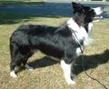 Border Collie
sannse 2003, WikiMedia Commons
 Boxer
BoxerLilly M 2007, WikiMedia Commons
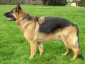 German Shepherd (Alsatian)
Elf 2004, WikiMedia Commons
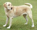 Retriever (Labrador)
Elf 2004, WikiMedia Commons


Clinical Signs
Signs depend on the severity of gingival hyperplasia and the degree to which the teeth are covered. They inclide pain on mastication, drooling +/- blood in saliva, Haemorrhage of the ginigiva, reluctance to eat and dysphagia. The animal may paw its mouth or rub its mouth along the floor.
Diagnosis
Clinical signs are indicative of the condition. A detailed oral examination under sedation will lead to a presumptive diagnosis.
Diagnostic Imaging
Oral radiographs should be taken to rule out concurrent conditions. One such condition is periodontitis which is demonstrated radiographically by alveolar bone loss associated with pocket formation between the tooth crown and gingiva.
Biopsy
Biopsy samples should include those areas of gingiva that show signs of inflammation with a softer than normal texture. Any gingiva with radiographic signs of bone involvement should also be sampled.
Treatment
The suspected cause of the condition should be corrected first. This may include a multimodal treatment plan aimed at controlling plaque formation including teeth brushing and providing the animal with sticks/toys that clean the teeth crowns.
Gingivectomy and gingivoplasty should be carried out under general anaesthetic if significant pseudo-pockets are present between the gingiva and teeth crowns. The aim should be to eliminate the pseudopockets and re-establish the normal anatomy of the gingival margin.
Electrosurgery and Laser surgery can be performed. Care must be taken with electrosurgery to avoid contact between the teeth crowns and the electrodes to prevent irreversible heat damage to the pulp.
Prognosis
The prognosis following surgical excision and histopathology is good. However, local recurrence is possible but less common if a treatment plan aimed at reducing plaque formation is implemented. A re-examination of the patient should be carried out at least every 6 months to assess for signs of recurrence and the sufficiency of plaque control measures.
References
Tutt, C., Deeprose, J. and Crossley, D. (2007) BSAVA Manual of Canine and Feline Dentistry (3rd Edition), British Small Animal Veterinary Association.
Merck & Co (2008) The Merck Veterinary Manual, Merial.