Difference between revisions of "Haemangiosarcoma"
| Line 26: | Line 26: | ||
==Laboratory Tests== | ==Laboratory Tests== | ||
====Haematology==== | ====Haematology==== | ||
| − | Anaemia will be evident which may be regenerative if due to blood loss, or microangiopathic due to the passage of red blood cells through the microvascular network of the tumour. | + | Anaemia will be evident which may be regenerative if due to blood loss, or microangiopathic due to the passage of [[erythrocytes|red blood cells]] through the microvascular network of the tumour. |
This results in the presence of schistocytes in dogs but not cats. | This results in the presence of schistocytes in dogs but not cats. | ||
A [[Neutrophilia|neutrophilia]] and [[Platelet Abnormalities#Thrombocytopaenia|thrombocytopenia]] may also be present. | A [[Neutrophilia|neutrophilia]] and [[Platelet Abnormalities#Thrombocytopaenia|thrombocytopenia]] may also be present. | ||
Revision as of 18:10, 18 August 2010
| This article is still under construction. |
Description
A highly malignant tumour of vascular endothelial origin. Commonly affect dogs and the most frequently affected areas are the spleen, pericardium, right atrium, liver and muscle. The cat is affected less frequently and the most common sites are the liver, spleen and mesentry. Metastasis occurs via the haematogenous route or via rupture and transabdominal spread. Metastatic sites include, lungs, liver, omentum and diaphram. Surgery is the treatment of choice but even with this survival time remains very short.
Signalment
Often found in German Shepherd Dogs and Golden Retrievers over 9 years of age. Domestic Short haired cats are the most commonly affected cat breed.
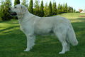 Golden Retriever
Monika Mężyńska (2005); WikiMedia Commons
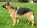 German Shepherd (Alsatian)
Ellen Levy Finch (22/02/04); WikiMedia Commons


Diagnosis
History and Clinical Signs
Can vary depending on the anatomic site that the mass is affecting. Signs such as anorexia, lethargy, weakness, vomiting and anaemia are common. Animals can also present Collapsed following the rupture of the mass, leading to a haemoabdomen or occasionally can be found dead.
In instances where the heart is involved animals may present in heart failure. If there is muscular involvement this often presents as a hard swollen mass.
If the nervous sytem is involved a range of neurological abnormalities will also be present.
Laboratory Tests
Haematology
Anaemia will be evident which may be regenerative if due to blood loss, or microangiopathic due to the passage of red blood cells through the microvascular network of the tumour. This results in the presence of schistocytes in dogs but not cats. A neutrophilia and thrombocytopenia may also be present.
Diagnostic Imaging
Radiography
Useful to look for evidence of metastasis.
Ultrasonography
This is sensitive in indentifying liver and splenic masses where the spleen will show a mixed or nonhomogenoeous pattern and the liver will look hypoechoic or anechoic. It can also be useful to detect metastatic spread.
Biopsy
The only way to to form a definitive diagnosis is following a biopsy and histopathology. This is needed to differentiate haemangiosarcoma from splenic haematoma, haemangioma and accessory splenic tissue.
Treatment
Surgery
Surgery is the treatment of choice for haemangiosarcoma in the dog and cat. All diseased tissue should be removed and Splenic haemangiosarcoma should be treated via splenectomy. Local removal is difficult if the pericardium and right atrium is involved. A pericardectomy can be undertaken but the prognosis with tumours at this location is grave.
Chemotherapy
This will provide a palliative treatment for animals with multiple masses or as an adjuvant therapy post-operatively. Doxorubicin based products are the most commonly used drugs for haemangiosarcomas.
Prognosis
Poor due to high risk of metastasis in the early course of the disease.
References
Ettinger, S.J. and Feldman, E. C. (2000) Textbook of Veterinary Internal Medicine Diseases of the Dog and Cat Volume 2 (Fifth Edition) W.B. Saunders Company.
Hall, E.J, Simpson, J.W. and Williams, D.A. (2005) BSAVA Manual of Canine and Feline Gastroenterology (2nd Edition) BSAVA
Nelson, R.W. and Couto, C.G. (2009) Small Animal Internal Medicine (Fourth Edition) Mosby Elsevier.