Difference between revisions of "Hepatitis, Chronic"
| Line 1: | Line 1: | ||
{{unfinished}} | {{unfinished}} | ||
| − | + | ==Introduction== | |
| − | + | '''Chronic hepatitis''' is an inflammatory-necrotising disease of at least 6 months duration. It is characterised by hepatocellular [[Adaptive Immunity to Viruses|apoptosis]] or [[Necrosis - Pathology|necrosis]], a variable mononuclear or mixed inflammatory cell infiltrate, [[Liver Regeneration|regeneration]] and [[Liver Fibrosis|fibrosis]] (1). It predominantly consists of lymphocytic-plasmacytic inflammatory infiltration, and the disease process typically involves a slowly progressive inflammation which leads to fibrosis and posibly cirrhosis. | |
| − | |||
==Signalment== | ==Signalment== | ||
*Common in dogs, especially young to middle-aged dogs. | *Common in dogs, especially young to middle-aged dogs. | ||
*Mixed and purebred dogs are affected but there is a familial predisposition in the following breeds: | *Mixed and purebred dogs are affected but there is a familial predisposition in the following breeds: | ||
| − | |||
<gallery> | <gallery> | ||
Image:Dobermann.jpg|'''Dobermann'''<p>John Adams (2007) WikiMedia Commons | Image:Dobermann.jpg|'''Dobermann'''<p>John Adams (2007) WikiMedia Commons | ||
| Line 20: | Line 18: | ||
Image:Beagle.jpg|'''Beagle'''<p> sannse (2003) WikiMedia Commons | Image:Beagle.jpg|'''Beagle'''<p> sannse (2003) WikiMedia Commons | ||
</gallery> | </gallery> | ||
| − | + | ==Aetiology== | |
| − | == | ||
| − | |||
| − | |||
A number of aetiologies include: | A number of aetiologies include: | ||
*Familial predisposition | *Familial predisposition | ||
| Line 32: | Line 27: | ||
*Autoimmune or steroid responsive disorder | *Autoimmune or steroid responsive disorder | ||
| − | |||
===Clinical Signs=== | ===Clinical Signs=== | ||
These include: | These include: | ||
| Line 41: | Line 35: | ||
*ascites - most consistent in dogs with [[Cirrhosis|cirrhosis]] | *ascites - most consistent in dogs with [[Cirrhosis|cirrhosis]] | ||
*and rarely [[Icterus|icterus]], seizures, fever and bleeding diathesis | *and rarely [[Icterus|icterus]], seizures, fever and bleeding diathesis | ||
| − | |||
===Laboratory tests=== | ===Laboratory tests=== | ||
| − | + | Haematology: | |
*Mild non-regenerative anaemia and microcytosis | *Mild non-regenerative anaemia and microcytosis | ||
| − | + | Biochemistry: | |
*Increased alanine aminotransferase (ALT) and alkaline phosphatase (ALP). However these may not be increased if end-stage [[Cirrhosis|cirrhosis]] is reached. | *Increased alanine aminotransferase (ALT) and alkaline phosphatase (ALP). However these may not be increased if end-stage [[Cirrhosis|cirrhosis]] is reached. | ||
*Hyperbilirubinaemia | *Hyperbilirubinaemia | ||
| Line 60: | Line 53: | ||
*Increased prolonged activated partial thromboplastin time (APTT) and prothrombin time (PT) indicates severe liver dysfunction or [[Disseminated Intravascular Coagulation|disseminated intravascular coagulation (DIC)]] | *Increased prolonged activated partial thromboplastin time (APTT) and prothrombin time (PT) indicates severe liver dysfunction or [[Disseminated Intravascular Coagulation|disseminated intravascular coagulation (DIC)]] | ||
| − | + | ==Imaging== | |
| − | |||
| − | |||
Abdominal radiographs will only reveal microhepatica or ascites when advanced stages of disease are reached. | Abdominal radiographs will only reveal microhepatica or ascites when advanced stages of disease are reached. | ||
| − | |||
Ultrasonographically, liver may be normal or non specific changes in echogenecity may be seen in early stages of the disease. In cases of [[Cirrhosis|cirrhosis]], microhepatica, irregularity in hepatic margin, focal lesions corresponding to regenerative nodules, hyperechogenicity of liver parenchyma associated with increased fibrous tissue and ascites may be seen. | Ultrasonographically, liver may be normal or non specific changes in echogenecity may be seen in early stages of the disease. In cases of [[Cirrhosis|cirrhosis]], microhepatica, irregularity in hepatic margin, focal lesions corresponding to regenerative nodules, hyperechogenicity of liver parenchyma associated with increased fibrous tissue and ascites may be seen. | ||
| − | + | ==Histopathology== | |
| − | |||
This is required for definitive diagnosis and to differentiate chronic hepatitis from other hepatopathies. Chronic hepatitis is characterised by moderate to severe lymphoplasmacellular [[Inflammation - Pathology|inflammation]] and [[Liver Necrosis|necrosis]] of the hepatocytes adjacent to the portal tracts. | This is required for definitive diagnosis and to differentiate chronic hepatitis from other hepatopathies. Chronic hepatitis is characterised by moderate to severe lymphoplasmacellular [[Inflammation - Pathology|inflammation]] and [[Liver Necrosis|necrosis]] of the hepatocytes adjacent to the portal tracts. | ||
| − | |||
==Treatment== | ==Treatment== | ||
| Line 85: | Line 73: | ||
**Penicillamine | **Penicillamine | ||
**Zinc | **Zinc | ||
| − | |||
==Prognosis== | ==Prognosis== | ||
Response to treatment is variable. Poor prognosis for dogs with [[Liver Fibrosis|fibrosis]] and [[Cirrhosis |cirrhosis]]. | Response to treatment is variable. Poor prognosis for dogs with [[Liver Fibrosis|fibrosis]] and [[Cirrhosis |cirrhosis]]. | ||
| − | |||
==References== | ==References== | ||
| Line 96: | Line 82: | ||
*Nelson, R.W. and Couto, C.G. (2009) '''Small Animal Internal Medicine (Fourth Edition)''' ''Mosby Elsevier''. | *Nelson, R.W. and Couto, C.G. (2009) '''Small Animal Internal Medicine (Fourth Edition)''' ''Mosby Elsevier''. | ||
| − | |||
| − | |||
| − | |||
| − | |||
[[Category:Liver_-_Inflammatory_Pathology]] | [[Category:Liver_-_Inflammatory_Pathology]] | ||
| − | |||
Revision as of 18:13, 1 November 2010
| This article is still under construction. |
Introduction
Chronic hepatitis is an inflammatory-necrotising disease of at least 6 months duration. It is characterised by hepatocellular apoptosis or necrosis, a variable mononuclear or mixed inflammatory cell infiltrate, regeneration and fibrosis (1). It predominantly consists of lymphocytic-plasmacytic inflammatory infiltration, and the disease process typically involves a slowly progressive inflammation which leads to fibrosis and posibly cirrhosis.
Signalment
- Common in dogs, especially young to middle-aged dogs.
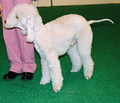
- Mixed and purebred dogs are affected but there is a familial predisposition in the following breeds:
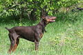 Dobermann
John Adams (2007) WikiMedia Commons
 Bedlington Terrier
Pleple2000 (2006) WikiMedia Commons
 Cocker Spaniel
Ellen Levy Finch (2004) WikiMedia Commons
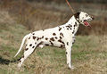 Dalmatian
Miroslav Cacik (2006) WikiMedia Commons
 Skye Terrier
Pleple2000 (2007) WikiMedia Commons
 Standard Poodle
John Leslie (2007) WikiMedia Commons
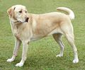 Labrador Retriever
Ellen Levy Finch (2004) WikiMedia Commons
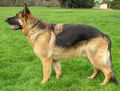 German Shepherd (Alsatian)
Ellen Levy Finch (2004) WikiMedia Commons
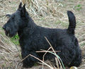 Scottish Terrier
Svencb (2003) WikiMedia Commons
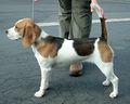 Beagle
sannse (2003) WikiMedia Commons


Aetiology
A number of aetiologies include:
- Familial predisposition
- Copper accumulation (copper storage disease)
- This may be a cause or consequence of chronic hepatitis. Copper is normally excreted in bile, therefore it can occur with any cholestatic hepatobiliary disorder.
- Chronic drug therapy
- Infectious, for example infectious canine hepatitis
- Autoimmune or steroid responsive disorder
Clinical Signs
These include:
- anorexia, lethargy and depression
- weight loss
- vomiting and diarrhoea
- polyuria and polydipsia
- ascites - most consistent in dogs with cirrhosis
- and rarely icterus, seizures, fever and bleeding diathesis
Laboratory tests
Haematology:
- Mild non-regenerative anaemia and microcytosis
Biochemistry:
- Increased alanine aminotransferase (ALT) and alkaline phosphatase (ALP). However these may not be increased if end-stage cirrhosis is reached.
- Hyperbilirubinaemia
- Hypoalbuminaemia
- Hyperglobulinaemia
- Decreased blood urea nitrogen (BUN)
- Hypoglycaemia
Further tests
- Increased bile acids
- Abnormal ammonia tolerance test
- Increased prolonged activated partial thromboplastin time (APTT) and prothrombin time (PT) indicates severe liver dysfunction or disseminated intravascular coagulation (DIC)
Imaging
Abdominal radiographs will only reveal microhepatica or ascites when advanced stages of disease are reached.
Ultrasonographically, liver may be normal or non specific changes in echogenecity may be seen in early stages of the disease. In cases of cirrhosis, microhepatica, irregularity in hepatic margin, focal lesions corresponding to regenerative nodules, hyperechogenicity of liver parenchyma associated with increased fibrous tissue and ascites may be seen.
Histopathology
This is required for definitive diagnosis and to differentiate chronic hepatitis from other hepatopathies. Chronic hepatitis is characterised by moderate to severe lymphoplasmacellular inflammation and necrosis of the hepatocytes adjacent to the portal tracts.
Treatment
- Glucocorticoids
- Taper down with improved clinical signs and normal liver enzymes values.
- This is not indicated for chronic hepatitis caused by drug therapy, primary copper accumulation or infectious agents.
- Response to treatment should be followed up by liver biopsy 3-6 months later as glucocorticoid causes steroid induced ALP
- Ursodeoxycholic acid at 15mg/kg PO SID
- It is a synthetic hydrophilic bile acid that has hepatoprotective (anti-inflammatory, immunomodulatory and antifibrotic effects) properties and choleretic effect. It expands the bile acid pool and displaces potentially hepatotoxic hydrophobic bile acids that accumulate in cholestasis.
- Vitamin E
- An antioxidant to scavenge free radicals which may contribute to oxidative hepatocellular injury.
- Copper chelation if copper exceeds 2000ppm
- Penicillamine
- Zinc
Prognosis
Response to treatment is variable. Poor prognosis for dogs with fibrosis and cirrhosis.
References
- (1) Van den Ingh, TSGAM et. al. (2006). Morphological classification of parenchymal disorders of the canine and feline liver. In Rothuizen J et. al., editors: WSAVA standards for clinical and histological diagnosis of canine and feline liver disease, Oxford, England, Saunders.
- Ettinger, S.J. and Feldman, E. C. (2000) Textbook of Veterinary Internal Medicine Diseases of the Dog and Cat Volume 2 (Fifth Edition) W.B. Saunders Company.
- Nelson, R.W. and Couto, C.G. (2009) Small Animal Internal Medicine (Fourth Edition) Mosby Elsevier.