Johne's Disease
| Also known as: | Paratuberculosis |
Johne's Disease is caused by Mycobacterium avium subsp. paratuberculosis. This chronic mycobacterial infection primarily affects the small intestine, and is most commonly seen in cattle, sheep and goats.
Johne's disease refers to the condition caused by the mycrobacterial infection. Calves are most commonly affected by this condition, and can be infected via a number of routes. They may be infected by ingestion of infected manure, or via transmammary infection. In addition, calves may also be infected in vitro, or may ingest the bacteria via colostrum.
There are three stages of infection in Johne's disease.
Stage 1: This often goes unnoticed, as it is subclinical. It typically affects animals less than two years of age, and will advance to stage II, only a few months later.
Stage 2: This is again a subclinical infection, usually affecting older heifers, or young adults. Infected animals are apparently healthy, but are shedding infection within their manure, infecting the environment.
Stage 3: Advanced infection. Animals will show clear clinical signs, including weight loss, decreased milk production, and a severe reduction in appetite.
Animals with advanced stage 3 will appear gaunt, and the meat is no longer deemed fit for human consumption. Once the disease is present in the herd, it is ver difficult to get rid of it.
Clinical
The infection primarily affects the intestine, and causes thickening of the mucosal wall. This causes impaired function, affecting its absorptive ability. This may cause, severe emaciation, and chronic profuse diarrhoea.
Clinical signs may also develop in older cows during times of stress; around calving for example.
In advances cases the infected animal gradually fades away and dies over the course of months.
Pathogenesis
Organisms penetrate the M-cells of the Peyer's patches. Mycobacteria invade macrophages and cause a granulomatous inflammatory response. Death may result from: damage to the mucosa, not absorbing nutrients, or inflammatory loss of protein.
Gross Pathology
The infected host appears emaciated.
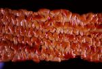
Fat is pale and oedematous, and only present in small amounts. Signs are confined to the terminal small intestine (especially the ileum) but are characteristic. It has diffusely thickened, velvety mucosa surface and transverse, corrugated ruggae with reddened crests. Infected animals may also have enlarged mesenteric lymph nodes
Changes are milder in sheep and goats, than seen in cows, and is symptoms are often missed.
Histologically
There are many large macrophages (epithelioid macrophages) in mucosa, submucosa and lymph nodes. The mesenteric lymph nodes are pale and enlarged (though not necrotic), and the lamina propria is infiltrated by sheets of macrophages with some lymphocytes. Acid-fast bacteria are found in the macrophages and giant cells, and this is detected by Ziehl-Neelson stain.
Bacteria act like foreign bodies producing a type IV hypersensitivity reaction. Sheep have two different forms: 1. Paucibacillary; many T cells, and few bacilli.
2. Multibacillary: Many macrophages, many bacilli in macrophages, and few lymphocytes.
Diagnosis
Diagnosis is by; Histology, Serological tests, ELISA and AGID.
60% of cases have lesions in colon and rectum and can be diagnosed by rectal biopsy.
