Nephron Microscopic Anatomy
Jump to navigation
Jump to search
|
|
Glomerulus
- Made up of many parallel capillaries
- These capillaries do not connect to venules as with other capillaries
- Blood flows into these capillaries through a wide afferent arteriole and leaves through a narrower efferent arteriole
- The flow from the efferant arteriole enters the peritubular capillaries surrounding the Proximal Tubule
- This change in diameter maintains a high filtration pressure which is essential for filtration
- Also the blood entering the afferent arteriole is at very high pressure already as it from the renal artery
- The pressure actually forces molecules through the glomerular filtration barrier which is responsible for selectively filtering the blood forming the glomerular filtrate.
- As well as the the cells in the blood vessels the other component of the glomerulus are the mesangial cells:
- These give support to the glomerulus
- Maintain glomerular basal lamina
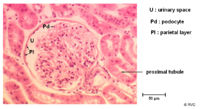
Bowmans Capsule
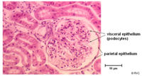
- Surrounds the capillaries of the glomerulus
- Has two layers
- Inner visceral layer - Podocytes
- Outer parietal layer
- It is here where the filtrate is collected before entering the proximal tubule
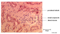
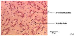
Proximal Tubule
- This is the piece of nephron which starts at the Bowmans capsule and ends in the loop of henle
- Consists of two parts which differ in cell morphology and function
- Pars convoluter - joins the urinary pole of the Bowmans capsule
- Pars recta (straight part) - links the pars convoluter to the descending thin limb of the loop of henle
- Has a brush border of densely packed microvilli to increase surface area
- The basal lamina is striated to increase the surface area for reabsorption
- Typical of actively ion transporting epithelial cells
- Lots of mitochondria to provide energy for exchange
- Lots of Na+ / K+ ATPases to maintain ion levels in cells at right level to allow efficient take up of ions from lumen
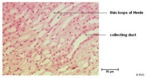
The Loop of Henle
The loop of henle basically consists of two parallel limbs which descend from the cortex into the medulla. They are joined at the bottom and as such the flow moves down one limb and up the other in opposite directions. This sets up a counter current exchanger and allows the loop of henle to be the major site of water reabsorption along the nephron. It has three parts; the thin decsending limb, the thin ascending limb and the thick ascending limb
