Difference between revisions of "Perineal Hernia"
JamesSwann (talk | contribs) |
JamesSwann (talk | contribs) |
||
| Line 1: | Line 1: | ||
{{unfinished}} | {{unfinished}} | ||
| − | |||
| − | |||
| + | ==Description== | ||
| + | '''Perineal hernia''' is the protrusion of pelvic or abdominal viscera through the structures of the weakened or ruptured pelvic diaphragm, causing displacement of the perineal skin. The pelvic diaphragm is composed of the coccygeus, levator ani and external anal sphincter muscles and, due to its proximity to these structures, the rectum is the organ most commonly involved in perineal hernias. Perineal hernias can be unilateral or bilateral and, depending of their location, they can be further classified as dorsal, ventral, sciatic or caudal hernias (see table). The herniated content is contained by the perineal fascia and, since the parietal peritoneum is usually intact, this constitutes a true [[Hernia|hernial sac]]. The sac may contain pelvic or retroperitoneal fat, peritoneal fluid, a deviation, dilation or sacculation of the rectum, a rectal diverticulum, the prostate gland, the urinary bladder (which may be retroflexed) or the small intestine and any of these organs may become [[Hernia|incarcerated]] or [[Hernia|stangulated]]. In cats, the sac frequently only contains the rectum. | ||
| + | |||
| + | The cause of perineal hernia is not know but several factors have been discussed, all of which are thought to contribute to progressive weakness and atrophy of the muscles of the pelvic diaphragm. These include: | ||
| + | *'''Breed predisposition''', with Welsh Corgis, Boston terriers, Boxers and Collies at particular risk. | ||
| + | *'''Sex''', with females at decreased risk of developing the condition as they have stronger pelvic diaphragm muscles. In dogs, 93% cases occur in intact males but the condition is more common in neutered male cats than entire males. | ||
| + | *'''Structural weakness of the pelvic diaphragm''' due to prolonged '''tenesmus''' which stretches the caudal rectal nerve and produces neurogenic atrophy of the muscles of the pelvic diaphragm. | ||
| + | *'''Tail docking''' may reduce the size and tone of the pelvic diaphragm muscles, increasing the risk of perineal hernia. Short-tailed Corgis have a greater risk of developing the condition than those with long tails. | ||
| + | *'''Congenital or acquired myopathies''', such as muscular dystrophy and myositis. | ||
| + | *'''Hormonal imbalances''', particularly increases in androgens (or in androgen receptors on the pelvic diaphragm muscles) and relaxin receptors which most commonly occur in male entire dogs. | ||
| + | *'''Prostatic enlargement''' is not thought to be a cause of perineal hernia ''per se'' but it contribute to the latter condition as it often produces faecal and urinary tenesmus. | ||
==Signalment== | ==Signalment== | ||
| − | + | The condition is much more common in dogs than cats and, in dogs, almost all cases are described in intact males. Most cases occur in animals older than 5 years and the median age in cats and dogs is 10 years. The following breeds have been shown to be predisposed to perineal hernias: | |
| − | |||
| − | |||
| − | |||
| − | |||
<gallery> | <gallery> | ||
| Line 17: | Line 22: | ||
Image:Pekingese.jpg|'''Pekingese'''<p>WikiCommons | Image:Pekingese.jpg|'''Pekingese'''<p>WikiCommons | ||
</gallery> | </gallery> | ||
| − | |||
| − | |||
| − | |||
| − | |||
| − | |||
| − | |||
| − | |||
| − | |||
| − | |||
==Diagnosis== | ==Diagnosis== | ||
| + | There may be a history of chronic tenesmus or of another disease causing generalised muscle weakness. | ||
===Clinical Signs=== | ===Clinical Signs=== | ||
| − | *perineal | + | Signs can be divided into physical signs related to the presence of the hernia and signs which occur when herniated organs become strangulated or obstructed. Clinical signs may therefore include: |
| − | * | + | *'''Physical signs''' |
| − | *dyschezia, tenesmus | + | **Swelling of the perineal area, either unilaterally or bilaterally. |
| − | *rectal prolapse | + | **Caudal projection of the anus (if the hernia is bilateral). |
| − | * | + | **The rectum loses lateral support on the affected side(s) and, on digital rectal examination, the skin over the hernia can be elevated because the rectum is no longer contained within the pelvic cavity. |
| − | * | + | **Constipation/obstipation, dyschezia and tenesmus result from alterations to the normal contours of the rectum, including deviations or flexures, sacculations (unilateral dilations), bilateral dilations and pulsion diverticula. Continued tenesmus may worsen the extent of the hernia. |
| − | * | + | **Chronic tenesmus and bilateral loss of rectal support may result in rectal prolapse. |
| − | * | + | *Signs due to incarceration or strangulations of organs and necessitating emergency treatment |
| − | + | **Stranguria or anuria due to retroflexion of the urinary bladder into the hernia. | |
| − | + | **Vomiting due to small intestinal obstruction | |
| − | + | *Signs after correction of the hernia | |
| + | **Faecal incontinence occurs commonly due to disruption of the caudal rectal nerves supplying the external anal sphincter. | ||
===Laboratory Tests=== | ===Laboratory Tests=== | ||
| − | |||
| − | |||
| − | |||
====Biochemistry==== | ====Biochemistry==== | ||
| − | + | Parameters indicating urinary tract obstruction (as occurs with retroflexion of the bladder) may be detected, including: | |
| − | + | *Raised serum urea and creatinine concentrations | |
| − | + | *Raised serum potassium concentration (not usually clinically significant until 24 hours after obstruction occurs). | |
| − | |||
===Diagnostic Imaging=== | ===Diagnostic Imaging=== | ||
| − | + | This is not necessarily required but radiography or ultrasonography may be useful to determine whether or not the bladder, prostate or small intestine are within the hernial sac. A positive contrast cystogram can be performed to better define the location of the bladder. | |
| − | This is not necessarily required but | ||
| − | |||
| − | |||
| − | |||
| − | |||
==Treatment== | ==Treatment== | ||
| Line 68: | Line 57: | ||
==References== | ==References== | ||
| + | *Fossum TW (1997) '''Small Animal Surgery''' ''Mosby'' | ||
*Ettinger, S.J. and Feldman, E. C. (2000) '''Textbook of Veterinary Internal Medicine Diseases of the Dog and Cat Volume 2''' (Fifth Edition) ''W.B. Saunders Company''. | *Ettinger, S.J. and Feldman, E. C. (2000) '''Textbook of Veterinary Internal Medicine Diseases of the Dog and Cat Volume 2''' (Fifth Edition) ''W.B. Saunders Company''. | ||
*Hall, E.J, Simpson, J.W. and Williams, D.A. (2005) '''BSAVA Manual of Canine and Feline Gastroenterology (2nd Edition)''' ''BSAVA'' | *Hall, E.J, Simpson, J.W. and Williams, D.A. (2005) '''BSAVA Manual of Canine and Feline Gastroenterology (2nd Edition)''' ''BSAVA'' | ||
Revision as of 19:47, 7 July 2010
| This article is still under construction. |
Description
Perineal hernia is the protrusion of pelvic or abdominal viscera through the structures of the weakened or ruptured pelvic diaphragm, causing displacement of the perineal skin. The pelvic diaphragm is composed of the coccygeus, levator ani and external anal sphincter muscles and, due to its proximity to these structures, the rectum is the organ most commonly involved in perineal hernias. Perineal hernias can be unilateral or bilateral and, depending of their location, they can be further classified as dorsal, ventral, sciatic or caudal hernias (see table). The herniated content is contained by the perineal fascia and, since the parietal peritoneum is usually intact, this constitutes a true hernial sac. The sac may contain pelvic or retroperitoneal fat, peritoneal fluid, a deviation, dilation or sacculation of the rectum, a rectal diverticulum, the prostate gland, the urinary bladder (which may be retroflexed) or the small intestine and any of these organs may become incarcerated or stangulated. In cats, the sac frequently only contains the rectum.
The cause of perineal hernia is not know but several factors have been discussed, all of which are thought to contribute to progressive weakness and atrophy of the muscles of the pelvic diaphragm. These include:
- Breed predisposition, with Welsh Corgis, Boston terriers, Boxers and Collies at particular risk.
- Sex, with females at decreased risk of developing the condition as they have stronger pelvic diaphragm muscles. In dogs, 93% cases occur in intact males but the condition is more common in neutered male cats than entire males.
- Structural weakness of the pelvic diaphragm due to prolonged tenesmus which stretches the caudal rectal nerve and produces neurogenic atrophy of the muscles of the pelvic diaphragm.
- Tail docking may reduce the size and tone of the pelvic diaphragm muscles, increasing the risk of perineal hernia. Short-tailed Corgis have a greater risk of developing the condition than those with long tails.
- Congenital or acquired myopathies, such as muscular dystrophy and myositis.
- Hormonal imbalances, particularly increases in androgens (or in androgen receptors on the pelvic diaphragm muscles) and relaxin receptors which most commonly occur in male entire dogs.
- Prostatic enlargement is not thought to be a cause of perineal hernia per se but it contribute to the latter condition as it often produces faecal and urinary tenesmus.
Signalment
The condition is much more common in dogs than cats and, in dogs, almost all cases are described in intact males. Most cases occur in animals older than 5 years and the median age in cats and dogs is 10 years. The following breeds have been shown to be predisposed to perineal hernias:
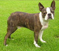 Boston Terrier
WikiCommons
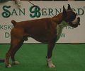 Boxer
Boxerdogsindepth.com
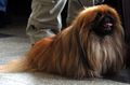 Pekingese
WikiCommons


Diagnosis
There may be a history of chronic tenesmus or of another disease causing generalised muscle weakness.
Clinical Signs
Signs can be divided into physical signs related to the presence of the hernia and signs which occur when herniated organs become strangulated or obstructed. Clinical signs may therefore include:
- Physical signs
- Swelling of the perineal area, either unilaterally or bilaterally.
- Caudal projection of the anus (if the hernia is bilateral).
- The rectum loses lateral support on the affected side(s) and, on digital rectal examination, the skin over the hernia can be elevated because the rectum is no longer contained within the pelvic cavity.
- Constipation/obstipation, dyschezia and tenesmus result from alterations to the normal contours of the rectum, including deviations or flexures, sacculations (unilateral dilations), bilateral dilations and pulsion diverticula. Continued tenesmus may worsen the extent of the hernia.
- Chronic tenesmus and bilateral loss of rectal support may result in rectal prolapse.
- Signs due to incarceration or strangulations of organs and necessitating emergency treatment
- Stranguria or anuria due to retroflexion of the urinary bladder into the hernia.
- Vomiting due to small intestinal obstruction
- Signs after correction of the hernia
- Faecal incontinence occurs commonly due to disruption of the caudal rectal nerves supplying the external anal sphincter.
Laboratory Tests
Biochemistry
Parameters indicating urinary tract obstruction (as occurs with retroflexion of the bladder) may be detected, including:
- Raised serum urea and creatinine concentrations
- Raised serum potassium concentration (not usually clinically significant until 24 hours after obstruction occurs).
Diagnostic Imaging
This is not necessarily required but radiography or ultrasonography may be useful to determine whether or not the bladder, prostate or small intestine are within the hernial sac. A positive contrast cystogram can be performed to better define the location of the bladder.
Treatment
Prognosis
References
- Fossum TW (1997) Small Animal Surgery Mosby
- Ettinger, S.J. and Feldman, E. C. (2000) Textbook of Veterinary Internal Medicine Diseases of the Dog and Cat Volume 2 (Fifth Edition) W.B. Saunders Company.
- Hall, E.J, Simpson, J.W. and Williams, D.A. (2005) BSAVA Manual of Canine and Feline Gastroenterology (2nd Edition) BSAVA
- Nelson, R.W. and Couto, C.G. (2009) Small Animal Internal Medicine (Fourth Edition) Mosby Elsevier.