Difference between revisions of "Small Animal Dermatology Q&A 10"
Jump to navigation
Jump to search
Ggaitskell (talk | contribs) |
|||
| Line 14: | Line 14: | ||
*These lesions also can be seen in non-Siamese kittens and will also resolve spontaneously. | *These lesions also can be seen in non-Siamese kittens and will also resolve spontaneously. | ||
*Adult cats of other breeds with widespread lesions should be evaluated carefully as these cats tend to have visceral involvement. | *Adult cats of other breeds with widespread lesions should be evaluated carefully as these cats tend to have visceral involvement. | ||
| − | |l1=Mast | + | |l1=Mast Cell Tumour |
|q2=What are the three major clinical presentations of MCT in cats? | |q2=What are the three major clinical presentations of MCT in cats? | ||
|a2= | |a2= | ||
| Line 27: | Line 27: | ||
*weight loss, and | *weight loss, and | ||
*anorexia. | *anorexia. | ||
| − | |l2= | + | |l2=Mast Cell Tumour |
|q3=What are the two histological subtypes of cutaneous MCT in cats? | |q3=What are the two histological subtypes of cutaneous MCT in cats? | ||
|a3= | |a3= | ||
| Line 33: | Line 33: | ||
#The histiocytic type occurs in young cats (<4 years of age) and is most common in Siamese cats. It frequently presents as subcutaneous nodules. Histologically, the mast cells are poorly granulated and lymphoid aggregates are common. Many of these tumors will spontaneously regress. | #The histiocytic type occurs in young cats (<4 years of age) and is most common in Siamese cats. It frequently presents as subcutaneous nodules. Histologically, the mast cells are poorly granulated and lymphoid aggregates are common. Many of these tumors will spontaneously regress. | ||
#The ‘mast cell form’ of cutaneous MCT tends to occur in mixed breed shorthaired cats. Lesions tend to be solitary and are discrete, nodular, papular, or plaque-like lesions in the dermis or subcutaneous tissue. It can also present as ‘miliary dermatitis’. | #The ‘mast cell form’ of cutaneous MCT tends to occur in mixed breed shorthaired cats. Lesions tend to be solitary and are discrete, nodular, papular, or plaque-like lesions in the dermis or subcutaneous tissue. It can also present as ‘miliary dermatitis’. | ||
| − | |l3= | + | |l3=Mast Cell Tumour |
</FlashCard> | </FlashCard> | ||
Latest revision as of 17:46, 3 September 2011
| This question was provided by Manson Publishing as part of the OVAL Project. See more small animal dermatological questions |
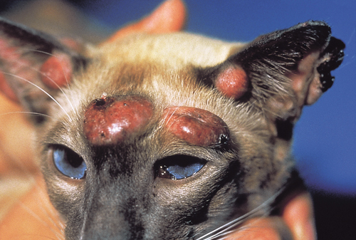
A 6-month old Siamese cat with multiple cutaneous nodules on its head, face, and ears is presented for examination. Skin biopsy findings reveal a histiocytic MCT.
| Question | Answer | Article | |
| What is the cat’s prognosis? |
|
Link to Article | |
| What are the three major clinical presentations of MCT in cats? | The three forms of clinical presentation are cutaneous, lymphoreticular or visceral, and gastrointestinal.
Clinical signs of lymphoreticular and gastrointestinal MCTs are indistinguishable:
|
Link to Article | |
| What are the two histological subtypes of cutaneous MCT in cats? | The two histological subtypes of feline mast cells are the histiocytic and mast cell type.
|
Link to Article | |
