Difference between revisions of "Small Animal Orthopaedics Q&A 01"
(Created page with "[[|centre|500px]] <br /> '''An intraoperative photograph of the trochlear ridges of the femoral condyle which have been exposed by a medial arthrotomy and lateral dislocation o...") |
|||
| (2 intermediate revisions by 2 users not shown) | |||
| Line 1: | Line 1: | ||
| − | [[|centre|500px]] | + | {{Manson |
| + | |book = Small Animal Orthopaedics Q&A}} | ||
| + | |||
| + | [[File:SmAnOrth 01.jpg|centre|500px]] | ||
<br /> | <br /> | ||
| Line 11: | Line 14: | ||
|a1= | |a1= | ||
There is extensive osteophyte development along the abaxial surfaces of the trochlear ridges. | There is extensive osteophyte development along the abaxial surfaces of the trochlear ridges. | ||
| − | |l1= | + | |l1=Degenerative Joint Disease |
|q2=Describe, on a microstructural level, how these structures develop. | |q2=Describe, on a microstructural level, how these structures develop. | ||
|a2= | |a2= | ||
| Line 23: | Line 26: | ||
Once established, osteophyte expansion may also proceed by surface growth. The mineralized areas develop a trabecular architecture and often retain a cartilage surface. Mature osteophytes may become incorporated into the metaphyseal trabecular structure during remodeling. | Once established, osteophyte expansion may also proceed by surface growth. The mineralized areas develop a trabecular architecture and often retain a cartilage surface. Mature osteophytes may become incorporated into the metaphyseal trabecular structure during remodeling. | ||
| − | |l2= | + | |l2=Degenerative Joint Disease |
</FlashCard> | </FlashCard> | ||
Latest revision as of 22:16, 23 October 2011
| This question was provided by Manson Publishing as part of the OVAL Project. See more Small Animal Orthopaedics Q&A. |
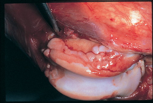
An intraoperative photograph of the trochlear ridges of the femoral condyle which have been exposed by a medial arthrotomy and lateral dislocation of the patella.
| Question | Answer | Article | |
| What are the pathologic structures present on the trochlear ridges? | There is extensive osteophyte development along the abaxial surfaces of the trochlear ridges. |
Link to Article | |
| Describe, on a microstructural level, how these structures develop. | Osteophytes develop in response to inflammation of the synovial tissues. Osteophytes develop primarily at the junction of the synovium and bone. When the synovium becomes inflamed, non-specific mutogenic factors are present within the fluid and the capsular tissues. Vascularity increases to the joint as a whole. Cells adjacent to bone proliferate and appear to be directed along the chondrogenic/osteogenic pathway of differentiation rather than the fibrogenic path, as occurs in the rest of the capsule. The fact that osteophytes are derived from tissue adjacent to bone or the proximity of bone matrix itself may be influencing factors. Continued presence of stimulatory factors leads to matrix deposition, mineralization and growth of osteophytes. Once established, osteophyte expansion may also proceed by surface growth. The mineralized areas develop a trabecular architecture and often retain a cartilage surface. Mature osteophytes may become incorporated into the metaphyseal trabecular structure during remodeling. |
Link to Article | |
