Difference between revisions of "Small Animal Soft Tissue Surgery Q&A 12"
Ggaitskell (talk | contribs) |
|||
| Line 21: | Line 21: | ||
Secondary inertia is caused by maternal factors (e.g. vulvar or pelvic obstruction; uterine torsion or rupture; | Secondary inertia is caused by maternal factors (e.g. vulvar or pelvic obstruction; uterine torsion or rupture; | ||
and uncommonly from hypocalcemia or hypoglycemia), and by fetal factors such as oversize fetuses (commonly with small litters and brachycephalic breeds), congenital disorders (e.g monster puppies and hydrocephalus), fetal malpositioning and dead fetuses. | and uncommonly from hypocalcemia or hypoglycemia), and by fetal factors such as oversize fetuses (commonly with small litters and brachycephalic breeds), congenital disorders (e.g monster puppies and hydrocephalus), fetal malpositioning and dead fetuses. | ||
| − | |l1= | + | |l1=Dystocia - Dog & Cat |
|q2=Does this dog have primary or secondary uterine inertia? | |q2=Does this dog have primary or secondary uterine inertia? | ||
|a2= | |a2= | ||
| Line 28: | Line 28: | ||
This dog has shown no clinical signs of active labor or whelping abnormality ruling out | This dog has shown no clinical signs of active labor or whelping abnormality ruling out | ||
secondary inertia as a problem and has not yet exceeded the gestational time range considered normal for ruling out primary uterine inertia. | secondary inertia as a problem and has not yet exceeded the gestational time range considered normal for ruling out primary uterine inertia. | ||
| − | |l2= | + | |l2=Dystocia - Dog & Cat |
|q3=What is the treatment, given your diagnosis? | |q3=What is the treatment, given your diagnosis? | ||
|a3= | |a3= | ||
| Line 38: | Line 38: | ||
After 69–70 days from the time of the second breeding (or ideally LH peak), primary uterine inertia is considered present, and diagnosis and treatment for dystocia initiated. | After 69–70 days from the time of the second breeding (or ideally LH peak), primary uterine inertia is considered present, and diagnosis and treatment for dystocia initiated. | ||
| − | |l3= | + | |l3=Dystocia - Dog & Cat |
</FlashCard> | </FlashCard> | ||
Revision as of 16:09, 18 October 2011
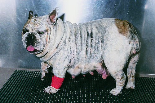
A three-year-old, female 84 English Bulldog is presented for suspected dystocia. It is 67 days since the first breeding and 65 days since the second. On examination the dog has marked mammary enlargement with minimal milk production, and a distended abdomen. No other abnormalities are noted.
| Question | Answer | Article | |
| Give several causes of dystocia. | Dystocia is considered when:
Causes include primary uterine inertia (lack of sufficient uterine contractions to expel a normal fetus from a normal birth canal) and secondary uterine inertia (lack of sufficient contractions because of exhaustion from prolonged labor or metabolic abnormality). Secondary inertia is caused by maternal factors (e.g. vulvar or pelvic obstruction; uterine torsion or rupture; and uncommonly from hypocalcemia or hypoglycemia), and by fetal factors such as oversize fetuses (commonly with small litters and brachycephalic breeds), congenital disorders (e.g monster puppies and hydrocephalus), fetal malpositioning and dead fetuses. |
Link to Article | |
| Does this dog have primary or secondary uterine inertia? | This dog does not have dystocia at this time. While the average bitch whelps around 63 days from the time of breeding, gestational length is variable ranging from 56–72 days. This dog has shown no clinical signs of active labor or whelping abnormality ruling out secondary inertia as a problem and has not yet exceeded the gestational time range considered normal for ruling out primary uterine inertia. |
Link to Article | |
| What is the treatment, given your diagnosis? | Treatment is nothing more than close observation over the next few days for signs of whelping. Rectal temperature is monitored: less than 37.7°C (100°F) indicates declining serum progesterone concentration and impending parturition (usually within 24 hours). If fetal viability is suspect, radiographs or sonography are performed. After 69–70 days from the time of the second breeding (or ideally LH peak), primary uterine inertia is considered present, and diagnosis and treatment for dystocia initiated. |
Link to Article | |
