Difference between revisions of "Snake Cardiovascular System"
Jump to navigation
Jump to search
(New page: {{unfinished}} The cardiovascular system of snakes is similar to other non-crocodilian reptiles but is modified for their linear shape. *'''Systemic venous return''' - Snakes have both re...) |
|||
| Line 4: | Line 4: | ||
*'''Systemic venous return''' - Snakes have both renal and hepatic portal circlations. Jugulars are located anterior to the heart near the trachea and may be cannulated by a cutdown procedure. The right jugular is larger than the left. Blood returns to the heart from the systemic circulation through the sinus venosus. | *'''Systemic venous return''' - Snakes have both renal and hepatic portal circlations. Jugulars are located anterior to the heart near the trachea and may be cannulated by a cutdown procedure. The right jugular is larger than the left. Blood returns to the heart from the systemic circulation through the sinus venosus. | ||
| + | [[Image:Burmese python heart in situ.jpg|200px|thumb|right|©RVC and its licensors, Peer Zwart and Fredric Frye. All rights reserved]] | ||
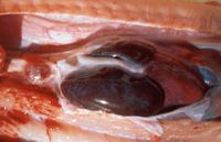
*'''Heart''' - The position of the heart varies among species and, as there is no diaphragm, it is mobile within the ribcage. This may facilitate the passage of prey. The heart has three chambers: right and left atria and one ventricle. The right atrium receives deoxygenated blood from the systemic circulation and the left receives oxygenated blood from lungs via the left and right pulmonary veins. The ventricle has internal ridges that enable a considerable functional separation between the oxygenated and deoxygenated blood. It is divided into three subchambers: the cavum pulmonale, cavum venosum and cavum arteriosum. | *'''Heart''' - The position of the heart varies among species and, as there is no diaphragm, it is mobile within the ribcage. This may facilitate the passage of prey. The heart has three chambers: right and left atria and one ventricle. The right atrium receives deoxygenated blood from the systemic circulation and the left receives oxygenated blood from lungs via the left and right pulmonary veins. The ventricle has internal ridges that enable a considerable functional separation between the oxygenated and deoxygenated blood. It is divided into three subchambers: the cavum pulmonale, cavum venosum and cavum arteriosum. | ||
*'''Systemic arteries''' - There are two aortae that leave the heart - the right aorta exits from the left side of the ventricle and the left aorta from the right side. The aortae join caudal to the heart to form the abdominal aorta that extends caudally through the coelomic cavity. Carotids extend cranially from the heart and adjacent to the trachea. | *'''Systemic arteries''' - There are two aortae that leave the heart - the right aorta exits from the left side of the ventricle and the left aorta from the right side. The aortae join caudal to the heart to form the abdominal aorta that extends caudally through the coelomic cavity. Carotids extend cranially from the heart and adjacent to the trachea. | ||
Revision as of 16:47, 25 February 2010
| This article is still under construction. |
The cardiovascular system of snakes is similar to other non-crocodilian reptiles but is modified for their linear shape.
- Systemic venous return - Snakes have both renal and hepatic portal circlations. Jugulars are located anterior to the heart near the trachea and may be cannulated by a cutdown procedure. The right jugular is larger than the left. Blood returns to the heart from the systemic circulation through the sinus venosus.
- Heart - The position of the heart varies among species and, as there is no diaphragm, it is mobile within the ribcage. This may facilitate the passage of prey. The heart has three chambers: right and left atria and one ventricle. The right atrium receives deoxygenated blood from the systemic circulation and the left receives oxygenated blood from lungs via the left and right pulmonary veins. The ventricle has internal ridges that enable a considerable functional separation between the oxygenated and deoxygenated blood. It is divided into three subchambers: the cavum pulmonale, cavum venosum and cavum arteriosum.
- Systemic arteries - There are two aortae that leave the heart - the right aorta exits from the left side of the ventricle and the left aorta from the right side. The aortae join caudal to the heart to form the abdominal aorta that extends caudally through the coelomic cavity. Carotids extend cranially from the heart and adjacent to the trachea.
