Difference between revisions of "Vascular Ring Anomalies"
| Line 1: | Line 1: | ||
| − | + | == Introduciton<br> == | |
| − | |||
| − | |||
| − | + | This congenital condition is most commonly found in dogs after weaning, but is very rare in cats. <br> | |
| − | + | Persistent Right Aortic Arch is the most common vascular ring anomaly, increased incidence in GSDs, and Irish setters. vascular ring forms between the ductus arteriosus/ligamentum arteriosum and the persistent right aorta. [[Megaoesophagus]] is seen cranial to the constriction. Other causes include a Double aortic arch and Anomalous subclavian arteries. These arise form the aortic arch rather than the brachiocephalic artery. | |
| − | [[Megaoesophagus]] is seen cranial to the constriction. | ||
| − | + | During normal embryonic development there are five pairs of aortic arch arteries (1-6, 5 is absent) that undergo developmental changes necessary to form the major arteries of the head, neck, and upper thorax. | |
| − | |||
| − | + | Malformations of the aortic arch arteries can cause several vascular ring anomalies, but the most commonly seen anomaly is persistent right aortic arch (PRAA). With PRAA, the esophagus and trachea are most often encircled by the left ligamentum arteriosum as they pass over the heart base causing chronic esophageal and tracheal compression. | |
| − | |||
| − | + | When a young animal with PRAA is weaned onto solid food, the esophageal and tracheal compression causes food stasis above the narrowed esophageal foramen leading to megaesophagus. Continued stress on the esophagus in this manner can cause permanent damage. Additionally, aspiration pneumonia and the resulting respiratory distress is a complication of postprandial regurgitation.<br> | |
| − | + | <br> | |
| − | |||
| − | |||
| − | + | == Clinical Signs<br> == | |
| − | |||
| − | |||
| + | Clinical signs are usually due to constriction of the oesophagus, such as regurgiation of food, usually noticed at weaning and aspiration pneumonia. This may ne seen as coughing. The animal may present as 'vomitting' but thorough physical exam should distinguish this from regurgitation. Respiratory distress may also be seen. Stunted growth, thin and malnourished with an increased appetite may also be a clinical sign. <br> | ||
| + | <br> | ||
| + | == Diagnosis''<br>'' == | ||
| + | History and clinical signs plus age and breed may be indicative of the disease. <br> | ||
| − | + | On physical exam, there will be no caridac murmours unless a PDA is alos present. A palpable dialted cervical oesophagus may be detected.<br> | |
| − | + | Radiographs should be taken for further diagnostics and will show a ventral deviation of the trachea and dilation of the oesophagus. A barium swallow will provide definative diagnosis of this condition.<br> | |
| − | + | <br> | |
| − | == | + | == Treatment''<br>'' == |
| − | + | Surgical resection of the vascular ring anomaly is possible, by refferal to a specialist cardiac surgeon. Dietary management is usually sufficient to control regurgitation, such as feeding from a platform and feeding small amounts of liquidised food. | |
| + | |||
| + | <br> | ||
<gallery> | <gallery> | ||
Image:GermanShep.jpg|'''German Shepherd (Alsatian)'''<p> Ellen Levy Finch (2004) WikiMedia Commons | Image:GermanShep.jpg|'''German Shepherd (Alsatian)'''<p> Ellen Levy Finch (2004) WikiMedia Commons | ||
Image:greatdane.jpg|'''Great Dane'''<p> Lilly M (2007) WikiMedia Commons | Image:greatdane.jpg|'''Great Dane'''<p> Lilly M (2007) WikiMedia Commons | ||
| − | </gallery> | + | </gallery> |
| − | |||
| − | |||
| − | |||
| − | |||
| − | |||
| − | |||
| − | |||
| − | |||
| − | |||
| − | |||
| − | |||
| − | |||
| − | |||
| − | |||
| − | |||
| − | |||
| − | |||
| − | |||
| − | |||
| − | |||
| − | |||
| − | |||
| − | |||
| − | |||
| − | |||
| − | |||
| − | |||
| − | |||
| − | |||
| − | |||
| − | |||
| − | |||
| − | |||
| − | |||
| − | |||
| − | |||
| − | |||
| − | |||
| − | |||
| − | |||
| − | |||
| − | |||
| − | |||
| − | |||
| − | |||
| − | |||
| − | |||
| − | |||
| − | |||
| − | |||
| − | + | <br> | |
| − | + | == Prognosis<br> == | |
| − | + | Good with surgery, but less favourable if there is extensive esophageal damage. If surgery cannot be performed, for what ever reason, then thoughts into the quality of life of the animal without surgery must be taken into consideration. | |
| − | [[Image:Praa.gif|right | + | [[Image:Praa.gif|thumb|right|125px|<center>Post-mortem specimen of an animal with a persistent right aortic arch <br /><small> Copyright Alun Williams 2009 (RVC))</center></small>]] <br> |
| − | |||
| − | + | == References<br> == | |
| − | + | Ettinger, S.J. and Feldman, E. C. (2000) Textbook of Veterinary Internal Medicine Diseases of the Dog and Cat Volume 2 (Fifth Edition) W.B. Saunders Company<br>Ettinger, S.J, Feldman, E.C. (2005) Textbook of Veterinary Internal Medicine (6th edition, volume 2)W.B. Saunders Company<br>Fossum, T. W. et. al. (2007) Small Animal Surgery (Third Edition) Mosby Elsevier <br>Merck & Co (2008) The Merck Veterinary Manual (Eighth Edition) Merial<br>Nelson, R.W. and Couto, C.G. (2009) Small Animal Internal Medicine (Fourth Edition) Mosby Elsevier. <br> | |
| − | + | <br> | |
| − | + | <br> | |
| + | == Test yourself with the Developmental Pathology Flashcards == | ||
| + | [[Cardiovascular Developmental Pathology Flashcards]] | ||
| − | [[Category:Cardiovascular_System_-_Developmental_Pathology]][[Category: | + | [[Category:Cardiovascular_System_-_Developmental_Pathology]] [[Category:Oesophagus_-_Pathology]] [[Category:To_Do_-_Review]] [[Category:Cardiac_Diseases_-_Cat]] [[Category:Oesophageal_Diseases_-_Cat]] [[Category:Cardiac_Diseases_-_Dog]] [[Category:Oesophageal_Diseases_-_Dog]] [[Category:Cardiac_Diseases_-_Horse]] [[Category:Oesophageal_Diseases_-_Horse]] [[Category:Cardiac_Diseases_-_Cattle]] [[Category:Cardiac_Diseases_-_Pig]] |
| − | [[Category:To_Do_- | ||
| − | [[Category: | ||
| − | [[Category: | ||
| − | [[Category: | ||
| − | [[Category: | ||
Revision as of 11:56, 15 March 2011
Introduciton
This congenital condition is most commonly found in dogs after weaning, but is very rare in cats.
Persistent Right Aortic Arch is the most common vascular ring anomaly, increased incidence in GSDs, and Irish setters. vascular ring forms between the ductus arteriosus/ligamentum arteriosum and the persistent right aorta. Megaoesophagus is seen cranial to the constriction. Other causes include a Double aortic arch and Anomalous subclavian arteries. These arise form the aortic arch rather than the brachiocephalic artery.
During normal embryonic development there are five pairs of aortic arch arteries (1-6, 5 is absent) that undergo developmental changes necessary to form the major arteries of the head, neck, and upper thorax.
Malformations of the aortic arch arteries can cause several vascular ring anomalies, but the most commonly seen anomaly is persistent right aortic arch (PRAA). With PRAA, the esophagus and trachea are most often encircled by the left ligamentum arteriosum as they pass over the heart base causing chronic esophageal and tracheal compression.
When a young animal with PRAA is weaned onto solid food, the esophageal and tracheal compression causes food stasis above the narrowed esophageal foramen leading to megaesophagus. Continued stress on the esophagus in this manner can cause permanent damage. Additionally, aspiration pneumonia and the resulting respiratory distress is a complication of postprandial regurgitation.
Clinical Signs
Clinical signs are usually due to constriction of the oesophagus, such as regurgiation of food, usually noticed at weaning and aspiration pneumonia. This may ne seen as coughing. The animal may present as 'vomitting' but thorough physical exam should distinguish this from regurgitation. Respiratory distress may also be seen. Stunted growth, thin and malnourished with an increased appetite may also be a clinical sign.
Diagnosis
History and clinical signs plus age and breed may be indicative of the disease.
On physical exam, there will be no caridac murmours unless a PDA is alos present. A palpable dialted cervical oesophagus may be detected.
Radiographs should be taken for further diagnostics and will show a ventral deviation of the trachea and dilation of the oesophagus. A barium swallow will provide definative diagnosis of this condition.
Treatment
Surgical resection of the vascular ring anomaly is possible, by refferal to a specialist cardiac surgeon. Dietary management is usually sufficient to control regurgitation, such as feeding from a platform and feeding small amounts of liquidised food.
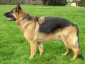 German Shepherd (Alsatian)
Ellen Levy Finch (2004) WikiMedia Commons
 Great Dane
Lilly M (2007) WikiMedia Commons


Prognosis
Good with surgery, but less favourable if there is extensive esophageal damage. If surgery cannot be performed, for what ever reason, then thoughts into the quality of life of the animal without surgery must be taken into consideration.

References
Ettinger, S.J. and Feldman, E. C. (2000) Textbook of Veterinary Internal Medicine Diseases of the Dog and Cat Volume 2 (Fifth Edition) W.B. Saunders Company
Ettinger, S.J, Feldman, E.C. (2005) Textbook of Veterinary Internal Medicine (6th edition, volume 2)W.B. Saunders Company
Fossum, T. W. et. al. (2007) Small Animal Surgery (Third Edition) Mosby Elsevier
Merck & Co (2008) The Merck Veterinary Manual (Eighth Edition) Merial
Nelson, R.W. and Couto, C.G. (2009) Small Animal Internal Medicine (Fourth Edition) Mosby Elsevier.