Canine Infectious Diseases: Self-Assessment Color Review, Q&A 02
| This question was provided by CRC Press. See more case-based flashcards |

|
Student tip: This case is a good beginning to the work-up of a case |
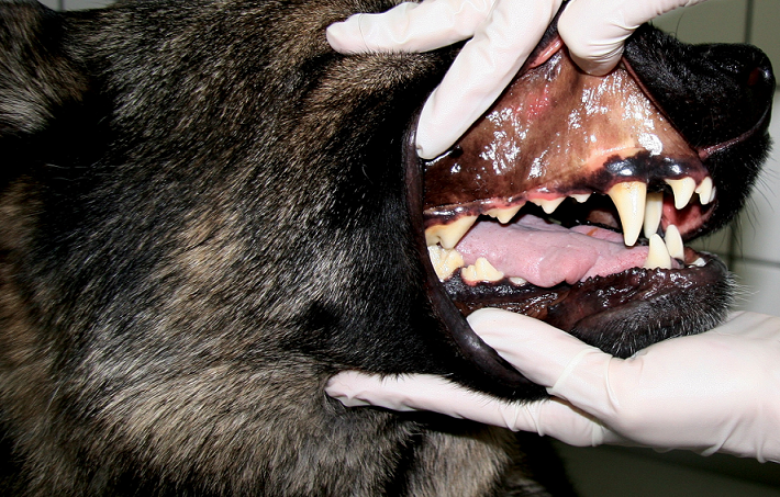
A 2-year-old intact male German Shepherd Dog mix was referred for a 4-day history of acute vomiting and anorexia. Two days before the dog was evaluated, the referring veterinarian reported icterus and had treated the dog with oral antibiotics (doxycycline) and a single injection of a short-acting glucocorticoid. Because the vomiting persisted, the owner did not administer the doxycycline. No previous health problems were known. The dog was up to date on vaccinations and regularly treated for endoparasites. He lived in a suburban area close to Munich, Germany, and had traveled to Southern Italy once, 10 months ago. On presentation, the dog was lethargic, but still responsive and ambulatory. He was in a good body condition (BCS 4.5/9). Rectal body temperature was 39.8°C (103.6°F), pulse was 120 bpm, and respiratory rate was 32 breaths/min. Mucous membranes were icteric (6). On palpation, the abdomen was tense, but not painful. The remainder of the physical examination was unremarkable.
| Question | Answer | Article | |
| What is the differential diagnosis for icterus? | Icterus is the description for yellowish pigmentation of skin and mucous membranes caused by hyperbilirubinemia. Hyperbilirubinemia can be classified into one of three categories of underlying etiologies: prehepatic (increased red blood cell destruction); hepatic (liver cell dysfunction); and posthepatic (a cholestatic disorder).
|
Link to Article | |
| What non-invasive tests would you perform first to investigate this problem? | First, a CBC should be performed. Moderate to severe regenerative anemia is most typical with prehepatic hyperbilirubinemia. A mild to moderate normocytic, normochromic, nonregenerative anemia is often present in chronic intra- and posthepatic inflammatory diseases (anemia of inflammatory disease) and should be discriminated from prehepatic hemolytic anemia. Changes in erythrocyte morphology consistent with increased erythrocyte destruction (e.g. spherocytes, schistocytes) are helpful to differentiate prehepatic from hepatic and posthepatic causes of anemia. Prehepatic hyperbilirubinemia is unlikely when the hematocrit is within the RI, without evidence of regeneration. A serum chemistry panel and urinalysis should be performed to determine whether there are liver function abnormalities (e.g. low cholesterol, albumin, glucose) that can indicate hepatic hyperbilirubinemia, or findings supportive of cholestatic disease (e.g. increased serum cholesterol concentration). The magnitude of serum liver enzyme activities can be influenced by a number of different factors, so these are more difficult to interpret. For example, both prehepatic (oxygen deficiency in hemolytic anemia) and posthepatic (e.g. bile duct obstruction due to pancreatitis) factors can cause secondary hepatocellular damage. An abdominal ultrasound is also recommended to aid differentiation of hepatic and posthepatic causes of hyperbilirubinemia. Posthepatic hyperbilirubinemia is unlikely when the gallbladder size, gallbladder wall thickness, diameter of the extrahepatic bile duct, and pancreas appear normal.
|
Link to Article | |
To purchase the full text with your 20% discount, go to the CRC Press Veterinary website and use code VET18.
