Cytology Q&A 01
| This question was provided by Manson Publishing as part of the OVAL Project. See more Cytology Q&A. |
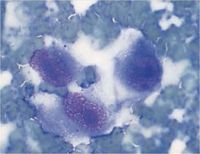
An eight-year-old Irish Setter presented with a mass in the right flank. An FNA was collected and a smear prepared.
| Question | Answer | Article | |
| What cell types and features are present in this photomicrograph (Wright–Giemsa, ×100 oil)? | Many erythrocytes and several cells with features of malignancy. These cells have large, round to oval nuclei with coarsely clumped chromatin and large, round to oval nucleoli. The cytoplasm is moderate and wispy to fusiform, with poorly defined borders. The rest of the smear had a similar appearance, with a few to a moderate number of cells with similar features. |
Link to Article | |
| What is your diagnosis? | Sarcoma. The features are not specific as to the type of sarcoma but the cytological features are considered to reflect an aggressive malignancy. The bloody nature of the aspirate may be due to contamination with blood, but the possibility of haemangiosarcoma should be considered when mesenchymal cells with features of malignancy are present. Evaluation for metastasis did not reveal any suspicious areas, so surgical removal was attempted. The histological diagnosis was haemangiosarcoma. |
Link to Article | |
