Question
Answer
Article
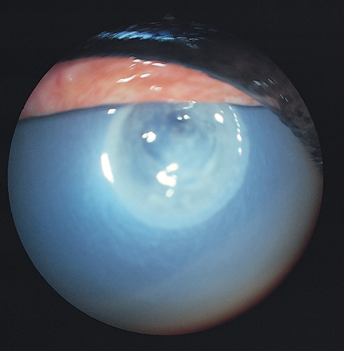
Is this ulcer infected or not?
Infected. There are
necrotic edges,
a cratered base,
diffuse corneal oedema,
severe injection and
severe pain. Link to Article
What is your most important diagnostic test?
A corneal scraping from the necrotic edges of this ulcer should be obtained for Gram staining and for culture.
Link to Article
If this ulcer was associated with a bacterial infection, how would you treat it?
Aggressive antibiotic therapy is indicated. Topical antibiotics should be applied frequently, preferably every hour initially.
This may be most effectively achieved using a subpalpebral lavage catheter.
After the first 12–24 hours, the rate of medication may be reduced to every 4–6 hours.
Selection of antibiotics should be based on the results of cytology, and confirmed by culture and sensitivity testing.
Topical atropine is indicated to relieve discomfort if miosis and ciliary spasm are present.
The ulcer should be carefully re-evaluated within 24 hours of initiation of treatment, and treatment modified as necessary. Link to Article
