Equine Peyer's Patches - Horse Anatomy
| This article is still under construction. |

Introduction
Peyer’s patches are lymphoid tissues found in the wall of the small intestine. They are part of the mucosal-associated lymphoid tissues (MALT) and more specifically the gut-associated lymphoid tissue (GALT). Although nodules of lymphatic tissue are found throughout the intestines in the small intestine, larger collections of nodules exist and these are referred to as Peyer’s patches. They may act as a primary lymphoid tissue in the horse.
Structure
Peyer’s patches are located in the lamina propria and submucosa of small intestine and may be distinguishable by the lack of villi covering them. The patches are regions of concentrated B lymphocyte follicles covered in a ‘dome’ of a specialised follicle associated epithelium (FAE) which consists of follicle associated enterocytes and M (microfold or multifold) cells.
M cells
M cells transport antigens from the intestinal lumen to the lymphocytes. Their luminal surface is folded and takes up antigens from the intestine via endocytosis and transports them to the extracellular space on their basal surface where the antigen is processed by antigen presenting cells.
Histology
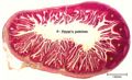
Intetsine w/Peyer's patch
©RVC 2008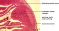
Peyer's patch
©RVC 2008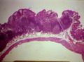
Peyer's patches
Courtesy of BioMed Archive


Function
Peyer’s patches have a role in maturation and differentiation of immature B lymphocytes. Antigens are presented to the B lymphocytes in the follicle which causes the B cells to become committed to IgA synthesis.