Gastric Ulceration - all species
Erosive and Ulcerative Gastritis
- Causes gastric ulcers
- Seen
- Commonly in the dog and pig.
- In young calves weaned onto a coarse diet.
- These usually heal as animal gets older.
- In the horse, associated with parasites.
- Once started, gastric ulcers can erode deeply.
- May penetrate gastric wall leading to peritonitis.
- May erode a blood vessel to cause haemorrhage.
Pathology
Gross
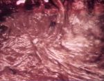
- Round or oval lesions from 1-4 cm in diameter.
- Sharply “punched out” lesions with perpendicular or slightly overhanging walls.
- Borders are level with, or slightly raised above, the surrounding mucosa.
- Depth is variable.
- Some penetrate the superficial mucosa only.
- Some deeply penetrate the muscularis externa.
- Base may be markedly haemorrhagic.
- In advanced chronic cases, scarring may result in a puckered appearance.
Histological
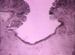
- Appearance varies with the degree aggressiveness of the ulcer and the amount of healing which has occurred.
- Rapidly excavating ulcers have minimal granulation tissue and collagen deposition.
- Others may have a necrotic base with a framework of granulation tissue and collagen.
- The blood vessels at the base of the ulcer may be thickened and thrombosed.
- In the bovine, the ulcer may have a superimposed fungal infection.
Pathogenesis
- There are differences in pathogenesis between species.

