Rheumatoid Arthritis
Jump to navigation
Jump to search
Pathogenesis:
- Type IV hypersensitivity: CD4 Th-1 cell mediated.
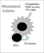
- Macrophages phagocytose self antigens and can present peptides on their MHC Class II molecules. The autoreactive TH-1 cells release IFN-gamma which activates macrophages to release prostaglandins, MMP enzymes and TNF-alpha (pro-inflammatory mediator), see diagram. These cytokines cause inflammation and tissue destruction.
- Occurs in the dog, mainly smaller breeds
- Uncommon
- Progressive erosive polyarthritis
- Mostly involves elbows, stifles, carpal and tarsal joints
- Grossly:
- Marked villus hypertrophy of synovial membrane
- Cartilage erosion
- Pannus and periarticular osteophyte formation
- In severe cases ankylosis
- Histologically:
- Hyperplasia of lining cells
- Proliferative synovitis
- Synovial membrane has fibrin deposits
- Lymphoid and plasma cell infiltration
- Surrounding haemorrhagic areas
- Macrophages containing haemosiderin
- Connective tissue may contain foci of necrosis
- Areas of erosion of peripheral articular cartilage and underlying subchondral bone
- Pathogenesis:
- May involve deposition of immune complexes within joints
- Substances degrading cartilage are released by synovial cells and macrophages involved in pannus formation
