Skin, superficial structures, conjunctiva and external ear
Four techniques are used to collect samples from the skin, subcutaneous masses and superficial structures; fine needle aspirates, swabs, impression smears and scrapings.
Fine needle aspirates
This technique is suitable for skin and subcutaneous masses including lymph nodes. Sedation or general anaesthesia is often not required but this depends on the nature of the patient and the location of the mass/lesion. Firm lesions may exhibit poor cellular exfoliation, often resulting in a small, non-representative sample or sampling haemorrhage. FNA is also used to assess fluid masses and cysts, however, the material aspirated may not be fully representative of underlying pathology since tissue cells are frequently absent.
Technique for solid masses
- Select a suitable needle. The length is selected in order to be inserted into all areas of the mass. Typically 23g, ¾ -1” for lymph nodes and large tissue masses and 25g for small lesions, attach a 5ml syringe. A ‘needle-only’ technique is also possible (see below)
- Local anaesthesia is usually not required and should be avoided if the lesion is superficial. Swab the skin with alcohol. Fix the lesion between thumb and forefinger and introduce the needle halfway into the lesion. Apply suction (0.5-1.0ml) by drawing back the plunger. Sample multiple sites in the lesion by advancing and retracting the needle without withdrawing the needle from the mass. The aspirate will often not be visible in the barrel of the syringe but sufficient cells will be present in the needle to prepare adequate smears
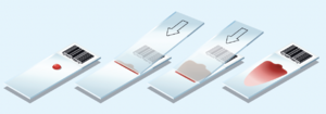
- Release the suction and withdraw the needle from the mass. Remove the needle, draw air into the syringe, re-attach, and carefully expel the sample onto two clean glass slides. Prepare smears using the same technique as for blood smears (Fig 1) and rapidly air dry. If the aspirate is too viscous for this technique, an alternative squash technique should be used (Fig 2) and rapidly air dried
- A ‘needle-only’ technique is also useful for superficial masses and lymph nodes. It may reduce haemodilution of the sample and lysis of fragile cells caused by suction. A 25g needle is inserted into the mass and redirected/retracted several times. Cells are collected by capillary action. The needle is withdrawn, a syringe containing air connected and the sample gently expelled onto slides. Smears are prepared as described above
Technique for fluid-filled cysts or masses
Collection of fluid from masses and cysts requires a 21-25g needle and a syringe. Swab the skin over the lesion with alcohol and aspirate 1-3ml of fluid. Prepare fresh smears as above and expel any remaining fluid into an EDTA and a plain tube. Add 2 drops of formalin to the plain tube and label accordingly. If an abscess is suspected then submit either a swab in transport medium or fluid in a sterile container for bacterial culture. If possible, perform FNA on any solid regions of the lesion and prepare air-dried smears.
Swabs
Smears made from swabs may be used for superficial skin lesions (pustules, vesicles, draining skin tracts) and conjunctival smears and are the method of choice for ear and vaginal cytology.
- Moisten a sterile cotton swab with sterile 0.9% NaCl. This may not be necessary in the case of exudative otitis externa
- Gently scrape or roll the swab across the lesion. An aural swab is usually obtained by passing the swab through an auroscope to sample the horizontal canal. Conjunctival swabs are obtained by first cleaning the conjunctival sac (sterile saline) and then inserting a swab into the conjunctiva and scraping to harvest cells. Topical anaesthesia is recommended
- Prepare two smears by gently rolling the swab along clean glass slides and rapidly air dry. Do not rub the swab across the slide as this will cause cell damage
Impression smears
Imprints are made from superficial skin lesions (scabs, ulcers, pustules, vesicles) for identification of organisms for example, Dermatophilus congolensis, Malassezia etc. Only superficial organisms/cells are sampled. The tape strip method is more commonly used for identification of Malassezia, particularly where skin folds or the local anatomy prevent good contact between the slide and the skin surface.
- Remove any scab material and gently touch a clean glass slide onto the surface of the lesion. If Dermatophilus congolensis infection is suspected then also imprint the underside of the scab
- Clean the lesion with a non-irritating antiseptic, wipe dry with sterile gauze and repeat the imprint
- Rapidly dry the smears and label ‘pre-cleaning’ and ‘post-cleaning
Scrapings
Scraping may occasionally be used to harvest cells from superficial or ulcerated skin lesions for example eosinophilic granuloma complex. It is the method of choice for conjunctival cytology.
- For skin lesions, gently scrape the surface with a sterile scalpel blade. For conjunctival scrapings, first clean the conjunctival sac with sterile saline then apply topical anaesthesia. Insert a flat, round tipped spatula or nylon bristle collection brush into the conjunctival sac and scrape 3 times in one direction to harvest cells from both the palpebral and the bulbar conjunctiva
- Deposit the scraping onto 2 clean glass slides and prepare squash smears (Fig 2). If a collection brush is used then cellular material should be rolled onto the slide
Sample collection summary
| Fine needle aspirate | Swab | Imprint | Scraping | |
|---|---|---|---|---|
| Conjunctiva | x | x | ||
| Ear | x | |||
| Lymph node | x | |||
| Skin lesion | x | x | x | x |
