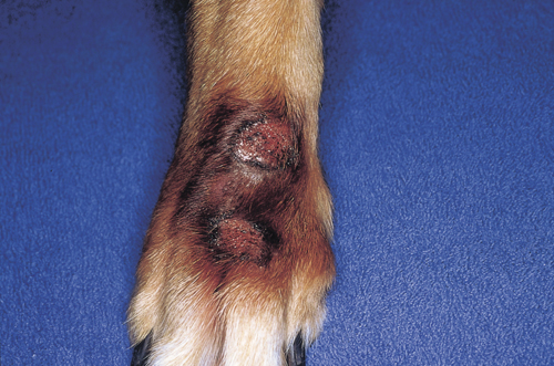
A 2-year-old dog was presented for evaluation of a mass on its forepaw. The lesion was raised, erythematous, moist, and firm to the touch. Closer examination revealed an erosive, circular lesion with a raised border; the lesion was slightly crater-like. A second healed lesion just distal to the first was also found, and extensive salivary staining was present on the paw. The owners reported the lesion had developed over the last several weeks, and this was the first occurrence of the lesion.
What are the two major causes of this syndrome?
What core diagnostic tests need to be done at this time?
What is the first line of therapy?
How would treatment proceed if the initial therapy failed?
The two major causes of this lesion are organic diseases and behavioral (obsessive–compulsive disorder). The latter is a diagnosis of exclusion, and this cannot be emphasized enough.
Pruritus in dogs is manifested by licking, and in many dogs this may be the only clue that the dog is pruritic. Sometimes the differentiation between the two major causes is obvious.
The dog has other compelling clinical signs of an underlying skin disease, there is a clear history of separation anxiety, or recent trauma/disruption in the dog’s life.
Clinical clues of an underlying pruritic skin disease may include signs of salivary staining on other limbs, a history of lesions developing on other legs in random fashion, and/or multiple lick granulomas occurring at the same time, or a history of trauma.
Initial diagnostic tests should include skin scrapings to rule out Demodex mites, impression smears to look for bacteria and/or yeast, and dermatophyte culture to rule out mycotoic infections, especially if the lesion is acute.
Assuming the skin scraping is negative, and while the fungal culture is pending, oral antibiotics, e.g. cephalexin (30 mg/kg PO q12h) would be the first choice therapy.
In the author’s experience, more than 75% of these lesions respond to antimicrobial therapy. This indicates that this lesion has an underlying trigger, particularly if the lesion responds completely but recurs at a later time.
Purely psychogenic lesions show only a minimal response to antibiotic therapy.
The most common cause of recurrent acral lick granulomas is atopy; atopic dogs often have multiple lick granulomas on different limbs, and owners report that the lesions shift from one site to another.
Lesions that do not respond to antimicrobial therapy should be biopsied to rule out
foreign body reactions,
kerion reactions,
neoplasia,
folliculitis, and/or
furunculosis.
Radiographs of the region should be taken looking for evidence of an underlying cause, e.g. fracture, osteosarcoma. The dog should be carefully examined for evidence of joint disease.
Atopy and/or food allergies should be investigated before making a definitive diagnosis of psychogenic dermatitis.
Tail dock neuroma is a rare complication of surgical tail docking. In this condition, the nerve endings regrow in a disorganized manner forming a neuroma.
Clinically, this appears as a swelling at the tail tip that stimulates pain or some other unpleasant sensation causing the dog to lick, chew, or mutilate the tail. Surgical removal is the treatment of choice.